Method and apparatus for ductal tube tracking imaging for breast cancer detection and diagnosis, and product

a technology of ductal tube and tracking imaging, which is applied in the field of breast cancer detection and imaging, can solve the problems of no imaging method has succeeded in tracking the full ductal system in vivo, and the architecture is very difficult to study in its entirety, so as to reduce the cost, shorten the time, and the effect of less expensiv

Active Publication Date: 2013-09-03
DDE MRI SOLUTIONS LTD
View PDF4 Cites 8 Cited by
  • Summary
  • Abstract
  • Description
  • Claims
  • Application Information

AI Technical Summary

Benefits of technology

[0027]The water movement inside the ductal trees is anisotropic, namely, water diffuses faster along the direction of the ducts (parallel to the ducts' walls) than in the orthogonal directions. The method of the present invention tracks anisotropic movement in the fibroglandular breast tissue using 3-dimensional diffusion tensor MRI, and thereby provides a means for mapping the ductal trees in the entire breast in vivo.
[0028]Moreover, since mammary malignancies originate in the epithelial tissues of the ducts, and spread inside the ducts, along ducts and consequently may outgrow the ducts, the invention postulates that the water diffusion in ducts with malignant growth will loose its anisotropic nature and will slow down due to the higher cell density as compared to the free water movements in the ducts. This change in the water diffusion characteristics can be detected by the method of the present invention using the above mentioned 3-dimensional diffusion tensor MRI and provide a means for the detection of breast cancer.
[0034]This is a new method for imaging the two breasts simultaneously to determine the breast ductal tubes and for detecting breast cancer. This method does not require injection of a contrast agent like the current leading MRI method for breast cancer detection. It is primarily directed towards developing MRI for screening to replace mammography screening. MRI has been applied previously to track fibers in the brain and in muscle tissue, but not in ductal breast tubes or other body tubes, which is a different application. The fact that a contrast agent is not necessary makes this inventive method purely non-invasive, non hazardous, less expensive and shorter in time. Consequently, the reduction of the costs and elimination of the discomfort and risk of contrast injection makes it much more practical for improving breast screening, particularly of high risk patients. By tracking the diffusion tensor in the ductal trees of the breast in vivo according to the present invention it should be possible to identify malignant growth in the ducts at early stages. The novel 3-dimensional magnetic resonance diffusion tensor imaging (MR-DTI) method for tracking the ductal systems (ductal trees), in the entire breast, based on sampling the water anisotropic diffusion along the mammary ductal network enables the foregoing. Any cancer growth in the ducts will modify this anisotropic water diffusion causing changes in both the diffusion direction and diffusion coefficient in each direction that will be detected by the method and apparatus of the present invention.
[0035]The invention provides an optimized protocol for high spatial resolution diffusion tensor imaging of the two breasts using echo-planar imaging (EPI) diffusion, and enables the presentation of final parametric images in the most revealing way for identifying breast abnormal tissue. The optimization of the protocol includes the diffusion weightings (b values), the number of non collinear directions (n≧6) for tensor calculations, the diffusion time and echo time, as well as the spatial resolution, signal to noise, and scanning time. Following calculation of the diffusion tensor, pixel by pixel, and diagonalizing the tensor to obtain the corresponding eigen values and eigenvectors per pixel reveals the anisotropy indices. Displaying the eigen values and their combination as parametric images and the prime eigenvector for vector presentation and ductal tracking shows the utility of the invention. The ability of DTI to track for the first time the ductal trees in the entire breast in vivo presents a revolutionized step forward and provides novel means for the detection, diagnosis and management of breast cancer patients.

Problems solved by technology

This architecture is very challenging to study in its entirety; thus far whole-breast lobe mapping has only been achieved for two human breasts using, ex-vivo, sliced tissue.
Currently, no imaging method has succeeded in tracking the full ductal system in vivo.

Method used


Image

  • Method and apparatus for ductal tube tracking imaging for breast cancer detection and diagnosis, and product
  • Method and apparatus for ductal tube tracking imaging for breast cancer detection and diagnosis, and product
  • Method and apparatus for ductal tube tracking imaging for breast cancer detection and diagnosis, and product

Examples


Embodiment Construction

High Level Flow Chart for DTI Mammary Ductal Tractography

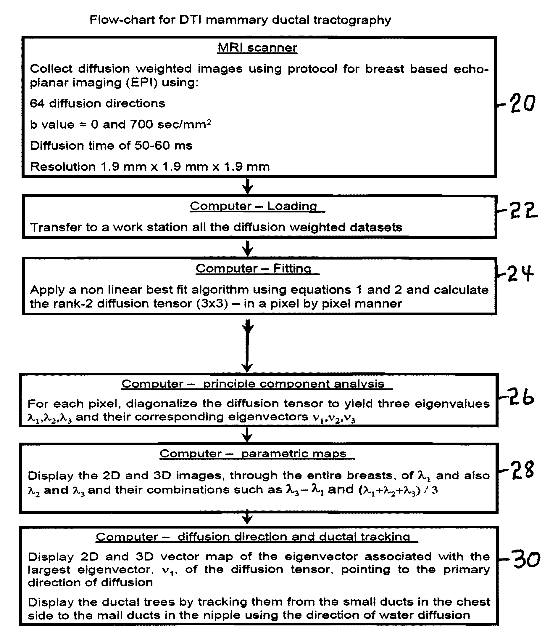
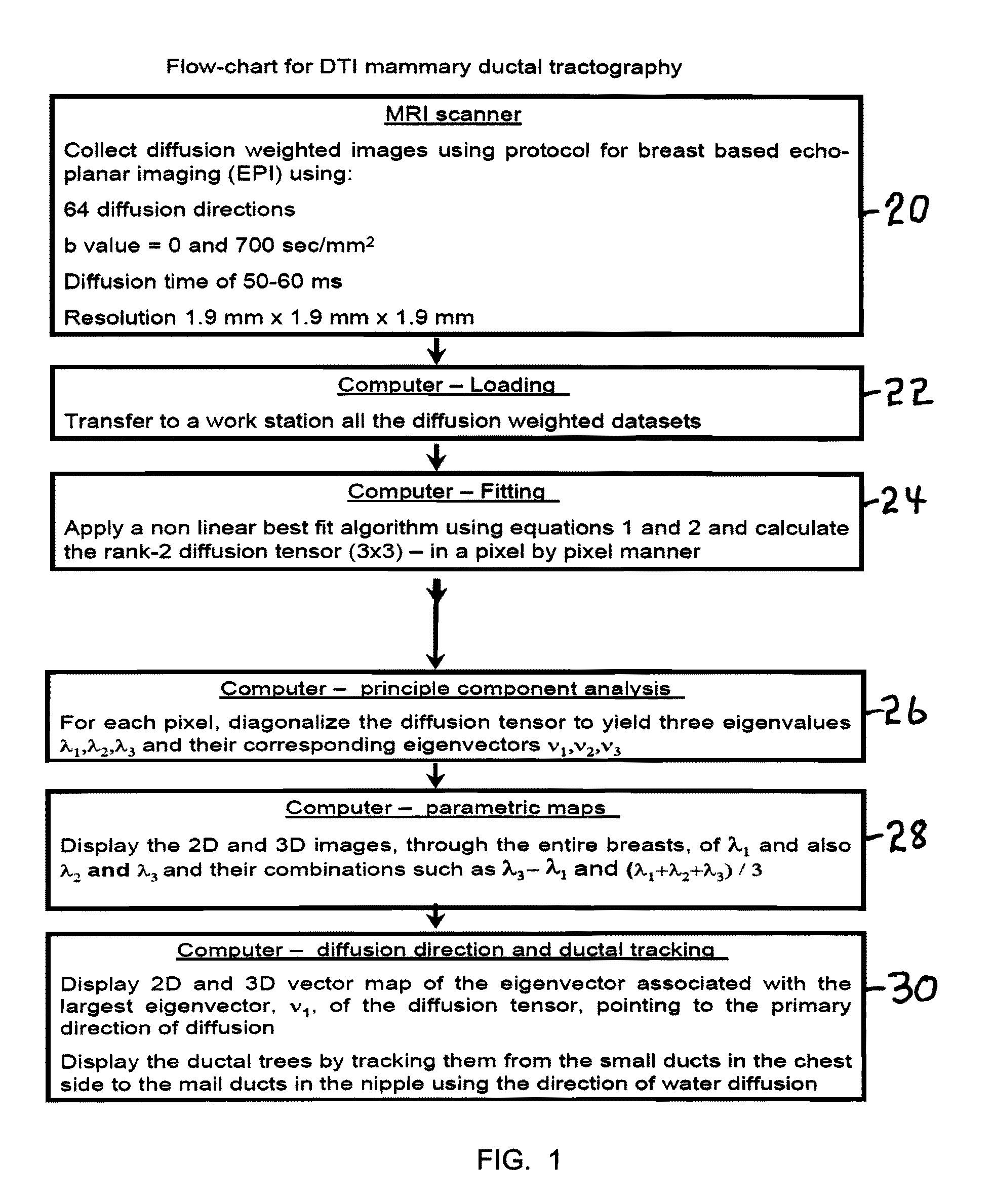
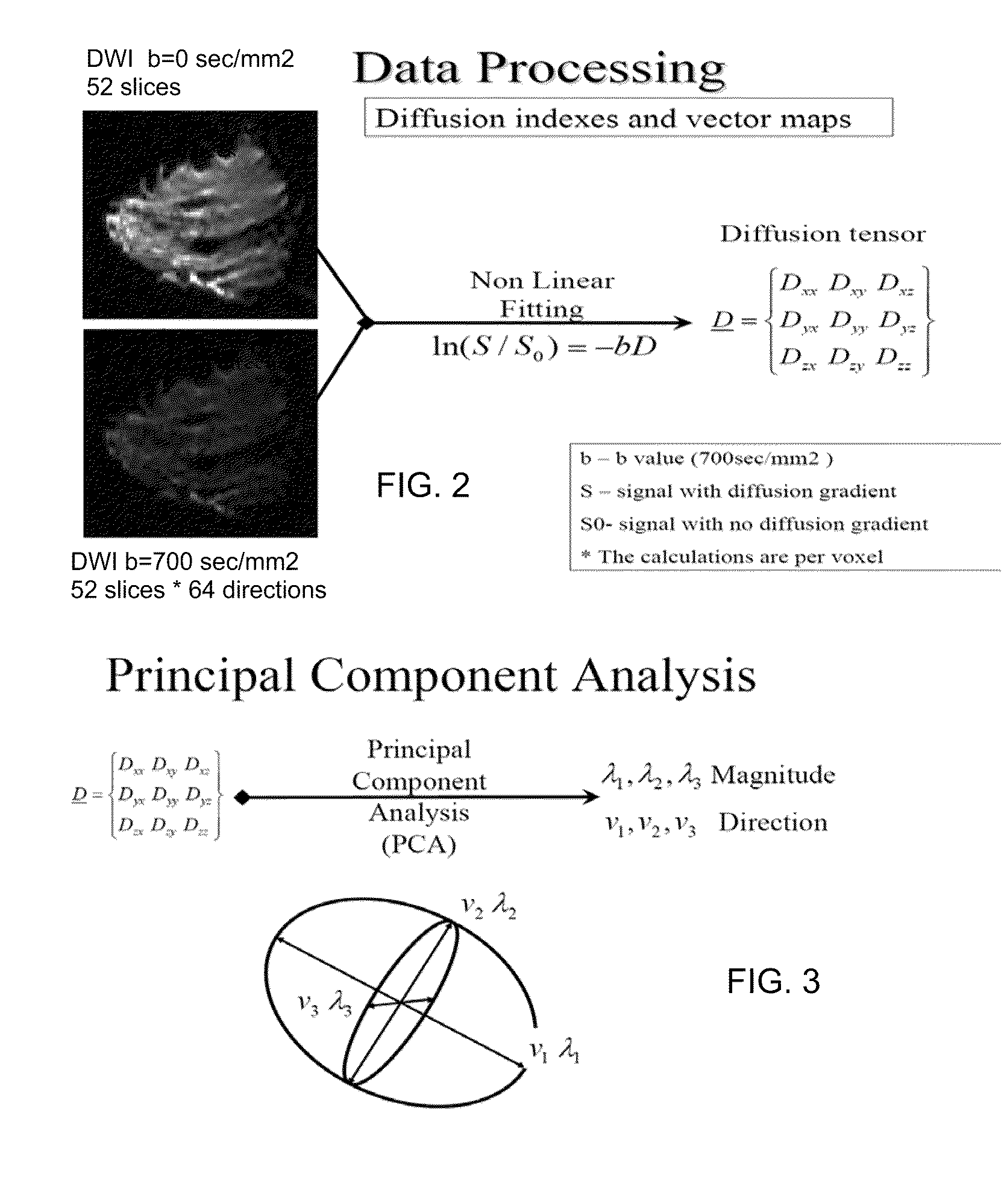
[0091]A flow chart of the novel method is shown in FIG. 1. In the first step 20, diffusion weighted images are collected using a protocol for breast based echo-planar imaging (EPI) using 64 diffusion directions, b value=0 and 700 sec / mm2, diffusion time of 50-60 ms and a resolution of 1.9 mm×1.9 mm×1.9 mm. The output of step 20 passes to step 22 Computer-Loading where all the diffusion weighted datasets are transferred to a work station. Next in step 24 Computer-Fitting a best fit algorithm is applied using equations 1 and 2 and the rank-2 diffusion tensor (3×3) is calculated in a pixel by pixel manner. The output of step 24 is passed to step 26 Computer-principle component analysis where for each pixel, the diffusion tensor is diagonalized to yield three eigenvalues λ1, λ2 and λ3 and their corresponding eigenvectors ν1, ν2 and ν3. The output of step 26 is passed to step 28 Computer-parametric maps to display the 2D and 3D ima...


PUM


Abstract

A method apparatus and computer product for imaging a human breast to map the breast ductal tree is disclosed. First, a breast is diffusion tensor imaged with high spatial resolution. Then the breast ductal tree is tracked using a protocol for breast based on echo-planar imaging (EPI) diffusion designed for optimizing diffusion weightings (b values), number of non-collinear directions for tensor calculations, diffusion, echo and repetition times, spatial resolution, signal to noise, scanning time and a sequence for fat suppression. The diffusion tensor is calculated by a non-linear best fit algorithm and then diagonalized with principal component analysis to three eigen vectors and their corresponding eigen values. A vector field map is obtained for tracking of breast ducts of the ductal trees along the direction of the 1st eigenvector v1 and the ductal tree is displayed on a voxel by voxel basis in parametric images using color coding and vector pointing.

Description

BACKGROUND OF THE INVENTION[0001]1. Field of the Invention[0002]The invention relates to a method for ductal tube tracking imaging for breast cancer detection and imaging, to an apparatus for ductal tube tracking, and to products comprising a computer readable medium comprising programs that includes displaying ductal tube images.[0003]2. Prior Art[0004]The two leading methods for screening the female population and detect breast cancer are currently high-quality X-ray mammography and breast ultrasound. The sensitivity of mammography has been estimated to be 63-88% and is lower by 10% or more for women aged 40-49 years and in older women using hormonal replacement therapy (HRT) than it is for other older women (Laya M B, Larson E B, Taplin S H, et al.: Effect of estrogen replacement therapy on the specificity and sensitivity of screening mammography. Journal of the National Cancer Institute 88(10): 643-649, 1996; and Persson I, Thurfjell E, Holmberg L: Effect of estrogen and estroge...

Claims


Application Information

Patent Timeline
Patent Type & Authority Patents(United States)
IPC IPC(8): G06K9/00
CPCG01R33/56341G01R33/5608
Inventor DEGANI, HADASSAEYAL, EREZ
Owner DDE MRI SOLUTIONS LTD
Why Eureka
Try Eureka