Suction and puncture device

A puncture needle and perforation technology, which is applied in the directions of puncture needle, inoculation and ovulation diagnosis, medical science, etc., to achieve the effect of reducing damage and damage, simple operation, and shortening the duration of puncture

Active Publication Date: 2014-10-22
KURUME UNIVERSITY +1
View PDF2 Cites 0 Cited by
  • Summary
  • Abstract
  • Description
  • Claims
  • Application Information

AI Technical Summary

Problems solved by technology

[0018] (2) In addition, in the above-mentioned puncture operation of 15 to 20 times, most of the samples cannot be extracted enough. In order to extract the desired amount of samples, Sometimes it is necessary to repeat the operation several times (usually three or four)
[0020] (4) In order to process the extracted sample and confirm whether the extraction is complete, etc., it is desired to perform cell diagnosis quickly, but because the extracted small cells are scattered , so it is difficult to use the naked eye to judge whether it can be extracted as a tissue, and it is difficult to extract in a sample shape that can meet the tissue diagnosis. Therefore, it is recommended that clinical laboratory technicians, pathologists, etc. be present, and there is a problem of labor and man-hours
[0023] (2) The puncture needle for Trucut on the market is an automatic type in which the outer cylinder is forcibly ejected from the tip when the button on the base end side is pressed. When the target organ is very hard, the ultrasonic endoscope may be pushed back to the hand due to the reaction force of the puncture
[0024] (3) It may be due to the complex structure of setting the inner needle in the outer cylinder and forming the tray part on the inner needle, compared with the suction method used Puncture needles are, for example, 10,000 to 30,000 yen, and puncture needles for Trucut are expensive, for example, around 50,000 yen
[0026] However, as mentioned above, the sample extracted by the aspiration method is the main pathological judgment method, and cytodiagnosis is the main method, although the probability is extremely low, but Tissue diagnosis can only be performed when a tissue sample that can satisfy the tissue diagnosis is extracted
Therefore, it cannot be said that it has fully functioned as a device for tissue diagnosis.
On the other hand, in the Trucut method, tissue diagnosis is the main purpose, but based on the above reasons, the scope of its application is limited by the size of the target organ and the position of the target organ that can be displayed by an ultrasonic endoscope. are limited, so the Trucut method is not widely used

Method used


Image

  • Suction and puncture device
  • Suction and puncture device
  • Suction and puncture device

Examples


Embodiment Construction

[0098] Hereinafter, embodiments of the present invention will be described based on the drawings.

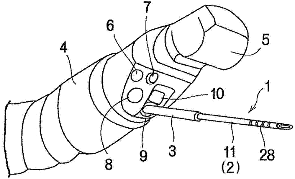
[0099] Such as figure 1 As shown, the suction and puncture device (1) of the present invention has a puncture needle (2), and the suction and puncture device (1) is mounted on an ultrasonic endoscope in a state covered by a protective tube (3). inside the mirror (4). A probe (5), a light guide (6), an air / water supply nozzle (7), an objective lens (8), a pliers mouth (9) and a pliers lifting platform (10) are arranged on the top end of the ultrasonic endoscope (4). , the above-mentioned puncture needle (2) is configured such that its tip can enter from the jaw of the forceps (9).

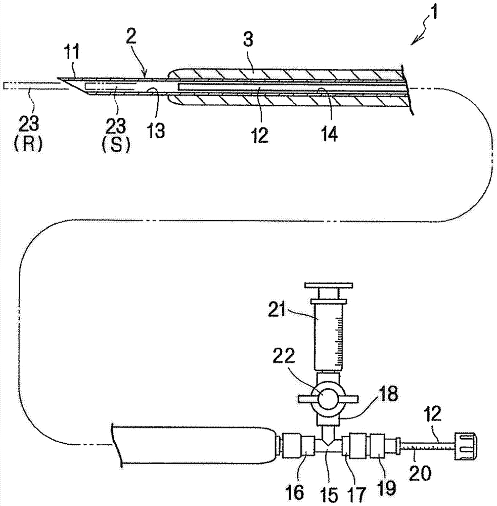
[0100] Such as figure 2 As shown, the above-mentioned puncture needle (2) has an outer cylinder (11) and a blocking member (12). Configure the top end. The opening edge of the top end of the outer cylinder (11) is formed in a knife shape. A sample storage portion (13) is formed between the top...


PUM


Abstract

A puncture needle (2) has an outer tube (11) that has an opening edge of a front end formed in the shape of a blade, and a stopper member (12) that has a front end disposed within the outer tube (11) at a position separated from the front end of the outer tube (11) by a predetermined distance. A sample containing portion (13) is formed between the front end of the stopper member (12) and the front end of the outer tube (11). A ventilation passage (14) is formed around the stopper member (12) within the outer tube (11). A suction device (21) is connected to the base end side of the outer tube (11). The suction device (21) is communicated and connected with the sample containing portion (13) via the ventilation passage (14).

Description

technical field [0001] The present invention relates to the collection of tissues, etc., from predetermined parts of organs or organs to be inspected in the body under the guidance of ultrasonic images, optical endoscopic images, or ultrasonic endoscopic images, etc. A suction and puncture method for examining a sample and a device used in the suction and puncture method, in more detail, relate to a method that can safely extract a tissue or the like at a predetermined site with a simple operation, and can extract any tissue without damaging the tissue. Aspiration and puncture method and aspiration and puncture device for large and small tissues. [0002] This application claims priority based on Japanese Patent Application No. 2010-89331 for which it applied in Japan on April 8, 2010, and uses the content here. Background technique [0003] Generally, in a method of extracting tissues, etc., from internal organs, etc., of an inspection target, there is a method of punctur...

Claims


Application Information

Patent Timeline
Patent Type & Authority Patents(China)
IPC IPC(8): A61B10/02A61B1/00
CPCA61B2017/306A61B10/0275A61B10/0283A61B1/018A61B17/3478A61B2010/045A61B1/00094A61B10/0266A61B17/3421A61B2017/320064
Inventor 冈部义信加治亮平石田祐介安元真希子山口知彦多比良朋希间宫朋彦柴木谦二
Owner KURUME UNIVERSITY
Why Eureka
Try Eureka