Automatic dividing method of ultrasound carotid artery vascular membrane

A technology of automatic segmentation and carotid artery, which is applied in the intersection of computer technology and medical images, and can solve problems such as many manual interventions.

Active Publication Date: 2014-12-31
HUAZHONG UNIV OF SCI & TECH
View PDF1 Cites 0 Cited by
  • Summary
  • Abstract
  • Description
  • Claims
  • Application Information

AI Technical Summary

Problems solved by technology

This method requires the operator to manually give the initial contour on each frame of the image, or give more initial points to form the initial contour, there is more manual intervention, and there is still a lot of room for improvement

Method used


Image

  • Automatic dividing method of ultrasound carotid artery vascular membrane
  • Automatic dividing method of ultrasound carotid artery vascular membrane
  • Automatic dividing method of ultrasound carotid artery vascular membrane

Examples


Embodiment Construction

[0054] The present invention will be further described in detail below in conjunction with the accompanying drawings and embodiments.

[0055] In the ultrasonic image provided by the invention, the computer automatic segmentation algorithm of the inner and outer contours of the carotid artery and plaques, its implementation steps are as follows:

[0056] (1) Determine the reference contour:

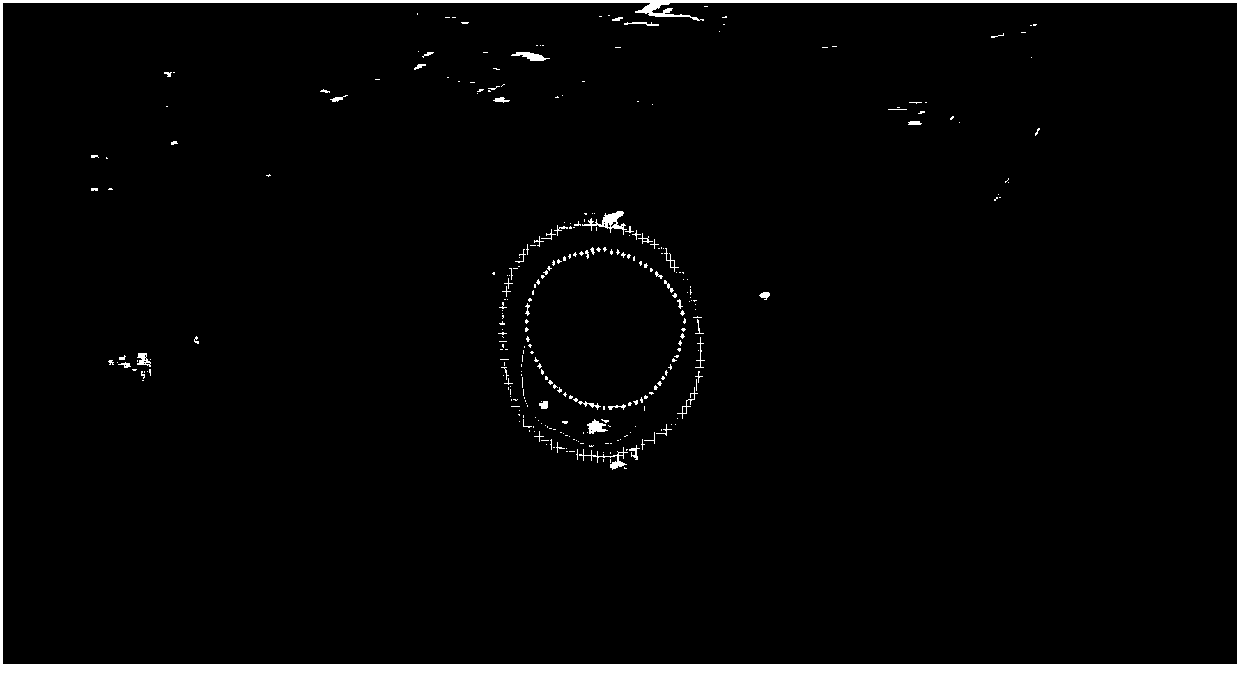
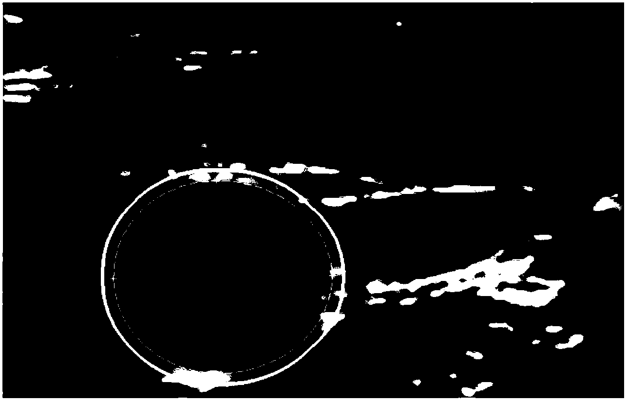
[0057] Load a group of carotid three-dimensional ultrasound volume data of a case into the computer, such as figure 2 shown. The computer automatically adjusts the voxel and volume according to the size of the voxel (mm 3 )proportion. The segmentation object of the present invention is each frame image of the three-dimensional ultrasound volume data of the carotid artery. If the current frame image is the first frame image of the three-dimensional ultrasound volume data of the carotid artery, the approximate position of the outer contour is judged empirically, and several obvious ref...


PUM


Abstract

The invention belongs to the field of intersection of a computer technology and medical images, and in particular relates to an automatic dividing method of internal and external profiles in the cross section direction of an ultrasound image carotid artery blood vessel. The method specifically comprises the following steps of: selecting the current frame image; manually defining a reference profile point and interpolating to form a reference profile; extrapolating the reference profile to form a first interested area; automatically detecting a carotid artery profile: searching a radial gradient maximum point on a polar coordinate image obtained through conversion in a first interested area as the initial external profile point, estimating the gray probability density distribution in the first interested area by using mixed distribution, carrying out level set evolution to obtain the external profile, interpolating the external profile to form an initial internal profile and carrying out level set evolution to obtain an internal profile. By adopting the method, ultrasound carotid artery images can be effectively divided, and the workload of doctors can be reduced; and besides, the carotid artery internal and external profiles obtained through division in the method can be used for calculating the size of the carotid artery blood vessel wall and providing accurate and effective information for research of carotid atherosclerosis.

Description

technical field [0001] The invention belongs to the intersecting field of computer technology and medical images, and in particular relates to an automatic segmentation method of intima and intima in the cross-sectional direction of a carotid artery in an ultrasonic image. Background technique [0002] Cardiovascular and cerebrovascular diseases are important diseases that endanger human health and cause death, such as myocardial infarction and stroke. The main cause of disease is carotid atherosclerosis. Compared with the traditional intima-media thickness (Intima-Media Thickness, IMT) measurement indicators, measurement indicators such as vessel wall volume (Vessel Wall Volume, VWV) can more accurately and intuitively reflect the status of atherosclerosis, so it has become a predictor of atherosclerosis in recent years. One of the important indicators of cardiovascular and cerebrovascular disease risk. [0003] Vessel wall volume can be measured on ultrasound carotid arte...

Claims


Application Information

Patent Timeline
Patent Type & Authority Patents(China)
IPC IPC(8): G06T7/00
Inventor 丁明跃李鹤程洁玉杨鑫
Owner HUAZHONG UNIV OF SCI & TECH
Why Eureka
Try Eureka