Optical coherence tomography (OCT) endoscope imaging device

Technology of an imaging device and endoscope

Inactive Publication Date: 2013-01-02
WUXI WIO TECH
View PDF7 Cites 17 Cited by
  • Summary
  • Abstract
  • Description
  • Claims
  • Application Information

AI Technical Summary

Problems solved by technology

[0002] The rigid endoscope system of the present invention has completely broken the working scope of traditional rigid endoscopes. Most of the core technologies of traditional rigid endoscopes use optical fiber bundles for light transmission and imaging, or use CCD technology for imaging. This type of endoscope It can only be observed from the surface of the tissue, so as to provide a basis for diagnosis for doctors. However, the symptoms of early cancer often occur at a depth of 1-3 mm below the epidermis, so the above-mentioned endoscope is powerless
In addition, there are also endoscopes that perform medical imaging through the principle of ultrasound. This type of endoscope can obtain tissue information deeper than the surface of biological tissues, but the resolution is only on the order of millimeters, and cannot effectively detect and diagnose fine tissues.

Method used


Image

  • Optical coherence tomography (OCT) endoscope imaging device
  • Optical coherence tomography (OCT) endoscope imaging device
  • Optical coherence tomography (OCT) endoscope imaging device

Examples


Embodiment Construction

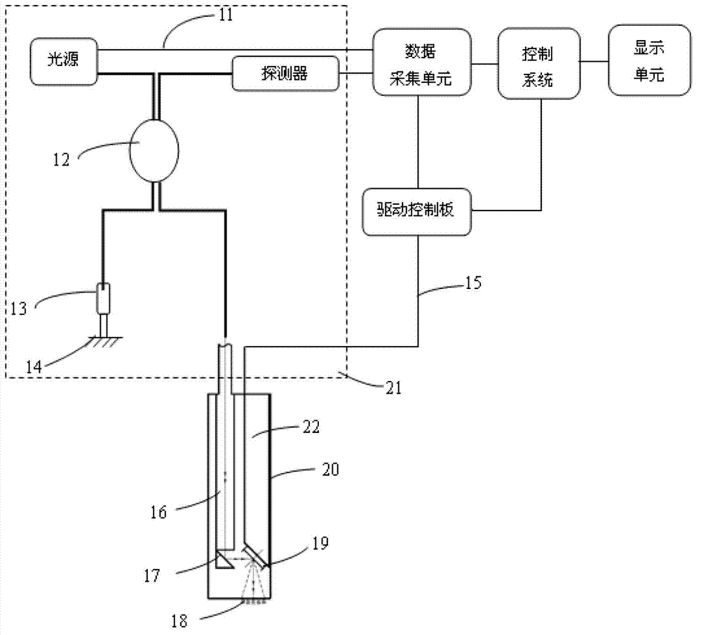
[0038] figure 1 As shown, it is an OCT endoscopic imaging device, including an endoscope with an OCT imaging system 21, and the OCT imaging system 21 includes a light source I, a light source II, a mixer 12, a detector, a reference arm and a sample arm The input end of the mixer 12 is respectively connected to the output ends of the light source I and the light source II through the optical fiber 11 or the spatial optical path, and its output end is respectively connected to the optical transmission ports of the reference arm and the sample arm through the optical fiber 11 or the spatial optical path; The input end of the detector is connected to the output end of the mixer 12 through an optical fiber 11 or a spatial optical path, and its output end is connected to the input end of the calculation control unit; the output end of the calculation control unit is connected to the input of the sample arm end. Wherein, the calculation control unit includes a data acquisition unit...


PUM


Abstract

The invention discloses an optical coherence tomography (OCT) endoscope imaging device. The OCT endoscope imaging device comprises an endoscope with an OCT imaging system, wherein the OCT imaging system comprises a light source I, a mixer, a probe, a reference arm and a sample arm; an input end of the mixer is connected with an output end of the light source I by an optical fiber or a space optical path; an output end of the mixer is connected to the reference arm and the sample arm by the optical fiber or the space optical path; an input end of the probe is connected to the output end of the mixer by the optical fiber or the space optical path; an output end of the probe and another output end of the light source I are respectively connected with an input end of a calculation control unit; and an output end of the calculation control unit is connected to an input end of the sample arm. By the endoscope device, nondestructive testing on viable tissues in the human body can be performed; and the device has micro-grade high resolution and is imaged in real time, can help doctors to diagnose the pathology in time, and is also used for industrial endoscope fields.

Description

technical field [0001] The invention relates to an optical image endoscope imaging device, in particular to an optical image endoscope based on the principle of optical coherence tomography (OCT). Background technique [0002] The rigid endoscope system of the present invention has completely broken the working scope of traditional rigid endoscopes. Most of the core technologies of traditional rigid endoscopes use optical fiber bundles for light transmission and imaging, or use CCD technology for imaging. This type of endoscope It can only be observed from the surface of the tissue, so as to provide diagnosis basis for doctors. However, the symptoms of early cancer often occur at a depth of 1-3 mm below the epidermis, so the above-mentioned endoscope is powerless. In addition, there are also endoscopes that perform medical imaging through the principle of ultrasound. This type of endoscope can obtain deeper tissue information below the surface of biological tissues, but the ...

Claims


Application Information

Patent Timeline
Patent Type & Authority Applications(China)
IPC IPC(8): A61B1/07A61B10/04A61B5/00
Inventor 傅霖来王东琳谢会开周正伟兰树明
Owner WUXI WIO TECH
Why Eureka
Try Eureka