
Diagnosis assistance apparatus

A technology for diagnosis assistance and judgment, which is applied in diagnosis, image analysis, medical science, etc., and can solve problems such as the inability to provide normal tissue and diseased tissue diagnosis

Active Publication Date: 2013-03-20
OLYMPUS CORP
View PDF11 Cites 3 Cited by
  • Summary
  • Abstract
  • Description
  • Claims
  • Application Information

AI Technical Summary

Problems solved by technology

[0005] However, the method of the above-mentioned conventional example is a method for detecting microcalcification points with high precision, and even if the running pattern of blood vessels and epithelial structures under the living body's submucosa is used, it cannot provide an effective method for diagnosis of normal tissue and diseased tissue. index

Method used


Image

  • Diagnosis assistance apparatus
  • Diagnosis assistance apparatus
  • Diagnosis assistance apparatus

Examples


no. 1 approach

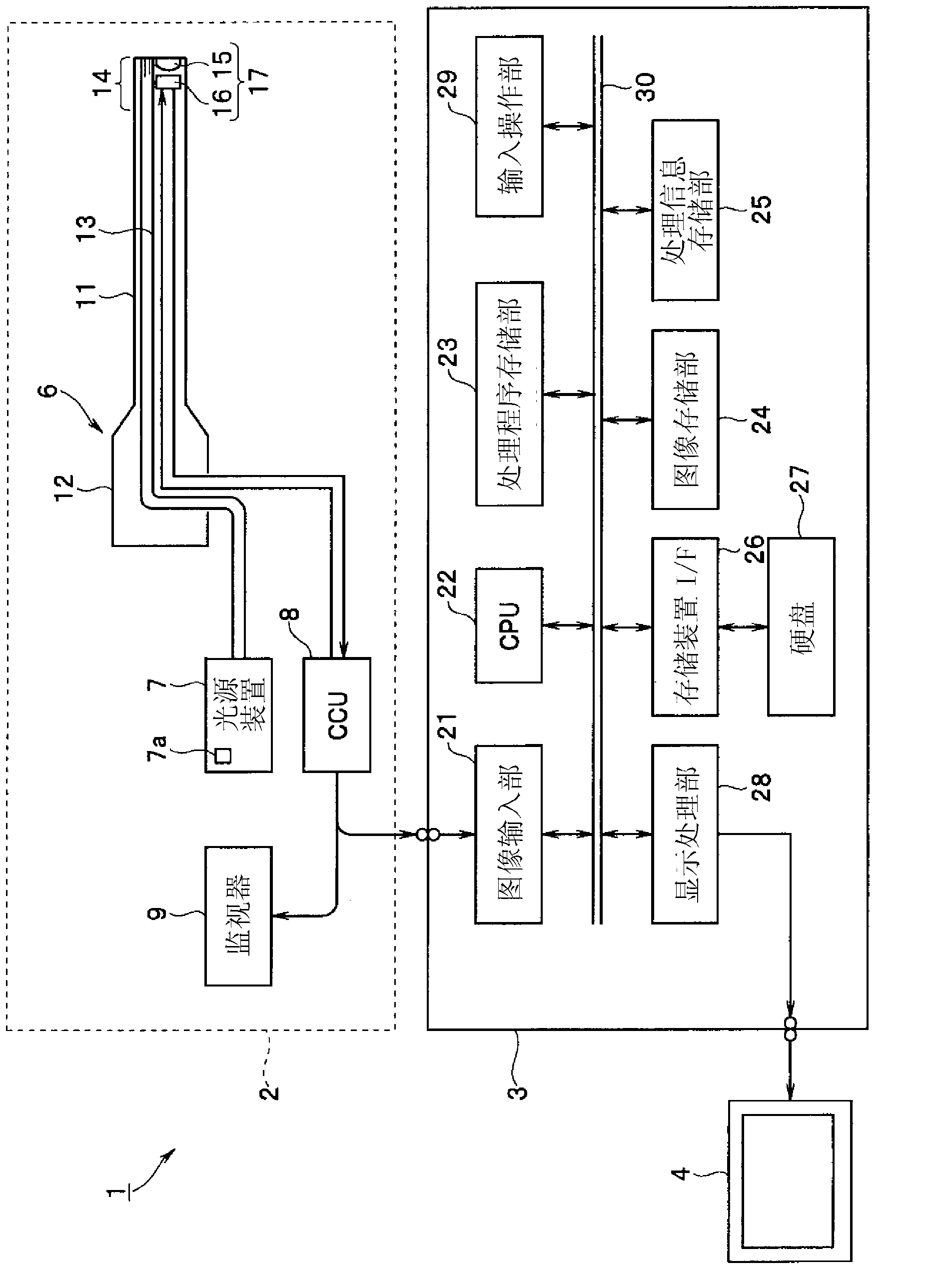
[0026] figure 1 The diagnostic support device 1 according to the first embodiment of the present invention shown includes an endoscope device 2; A personal computer for performing image processing on the mirror image;

[0027] The endoscope device 2 has: an endoscope 6, which is inserted into a body cavity to form an in-vivo imaging device for photographing the body; a light source device 7, which supplies illumination light to the endoscope 6; and a camera control unit (abbreviated as a signal processing device). A CCU) 8 processes signals from the imaging unit of the endoscope 6; and a monitor 9 displays an image captured by the imaging element as an endoscopic image by being input with an image signal output from the CCU 8.

[0028] The endoscope 6 has an insertion portion 11 inserted into a body cavity, and an operation portion 12 provided at the rear end of the insertion portion 11 . In addition, a light guide 13 that transmits illumination light penetrates inside the ...

no. 2 approach

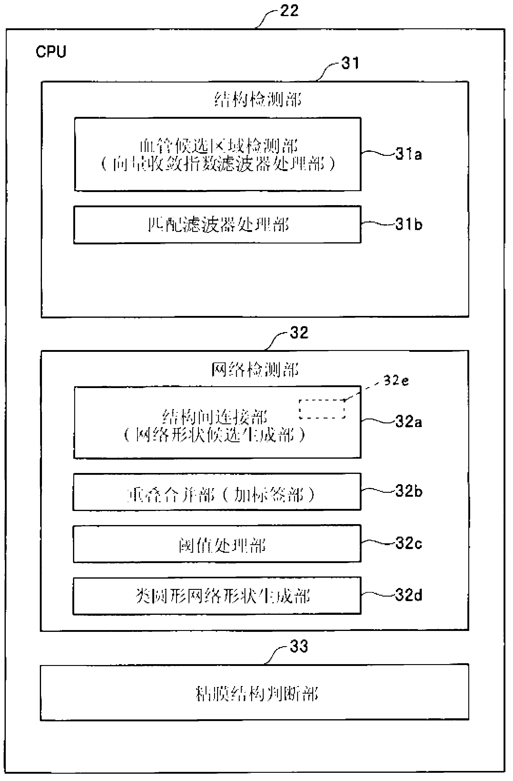
[0102] Next, a second embodiment of the present invention will be described. In the first embodiment, focusing on the running pattern of the blood vessel, a circular-like structure was detected in order to detect the structure of the running pattern of the blood vessel. Structure. And, it is used to assist the diagnosis of epithelial structure according to the detection results.

[0103] The structure on the hardware of this embodiment mode and figure 1 The first embodiment shown is the same, and this embodiment is the same as that in the first embodiment figure 2 The processing function of the structure detection part 31 of the CPU22 is slightly different. Figure 13 A block diagram showing processing functions of the CPU 22 in this embodiment.

[0104] Such as Figure 13 As shown, the CPU 22 has a structure detection unit 31, a network detection unit 32, and a mucosal structure determination unit 33, but is configured to use an epithelial candidate region detection un...


PUM


Abstract

The diagnosis assistance apparatus is provided with: an image input unit for inputting endoscopic images acquired from living tissue; a structure detection means for detecting elliptical structures from image signals that correspond to the endoscopic images input into the image input unit; and a network detection unit that detects a prescribed network topology in the endoscopic image on the basis of the number of elliptical structures detected by the structure detection unit, the area of elliptical structures, the distance between elliptical structures, or the number of connections between elliptical structures.

Description

technical field [0001] The present invention relates to a diagnosis assistance device for performing image processing for diagnosis assistance on endoscopic images of living tissue. Background technique [0002] In recent years, endoscopes capable of inserting an insertion portion into body cavities such as alimentary canals to image living mucous membranes such as the inner walls of alimentary canals and display them as endoscopic images have been widely used in the medical field. [0003] In addition, research has been conducted on image processing for auxiliary diagnosis using the running patterns of blood vessels and epithelium (or epithelial tissue) under the living body submucosa in endoscopic images captured by an endoscope. [0004] On the other hand, the conventional example of Japanese Unexamined Patent Publication No. 9-251535 discloses an image processing method in which a pixel is radiated radially from an arbitrary pixel to surrounding pixels on medical image d...

Claims


Application Information

Patent Timeline
IPC IPC(8): A61B1/00A61B1/04
CPCA61B1/05A61B1/00009G06T2207/30092G06T7/0012G06T7/00G06T2207/30096G06T2207/10068G06T2207/20148G06T7/136A61B1/000094
Inventor 田中健一
Owner OLYMPUS CORP
Learn More