Time-resolved multicolor single-energy X-ray imaging spectrometer

A time-resolved, X-ray technology, applied in the field of X-ray imaging, can solve the problems of limited channel number, only 2 channels, curved crystal imaging and insufficient spatial resolution of MMI, and achieve high spatial resolution and high light collection efficiency , the effect of high integral efficiency

Inactive Publication Date: 2013-08-07
TONGJI UNIV
View PDF5 Cites 12 Cited by
  • Summary
  • Abstract
  • Description
  • Claims
  • Application Information

AI Technical Summary

Problems solved by technology

[0004] The above three methods still have the following shortcomings under certain ICF physical experiment types and strong laser device energy conditions: 1. The spatial resolution of curved crystal imaging and MMI is insufficient, and it cannot achieve a spatial resolution better than 5 microns. Although GMXI has The spatial resolution is better than 5 microns, but the number of channels is only 2; 2. When performing X-ray self-luminescence imaging, the intensity of the self-luminescence continuum of the bremsstrahlung is much lower than the line spectrum of the characteristic radiation, especially at higher energy In the diagnostic area of ​​the segment (such as 2-5keV) or a strong laser device with only kJ level of energy (such as Shenguang II), this type of experiment uses a four-channel KB microscope with X-ray multilayer film elements to sacrifice a certain amount of energy Obtaining higher light collection efficiency at the expense of resolution is a feasible solution, but there is still the problem of limited number of channels
This process requires the images of multiple channels to be arranged along the same straight line within the length of the photocathode slit, and the above three methods cannot meet this requirement

Method used


Image

  • Time-resolved multicolor single-energy X-ray imaging spectrometer
  • Time-resolved multicolor single-energy X-ray imaging spectrometer
  • Time-resolved multicolor single-energy X-ray imaging spectrometer

Examples


Embodiment 1

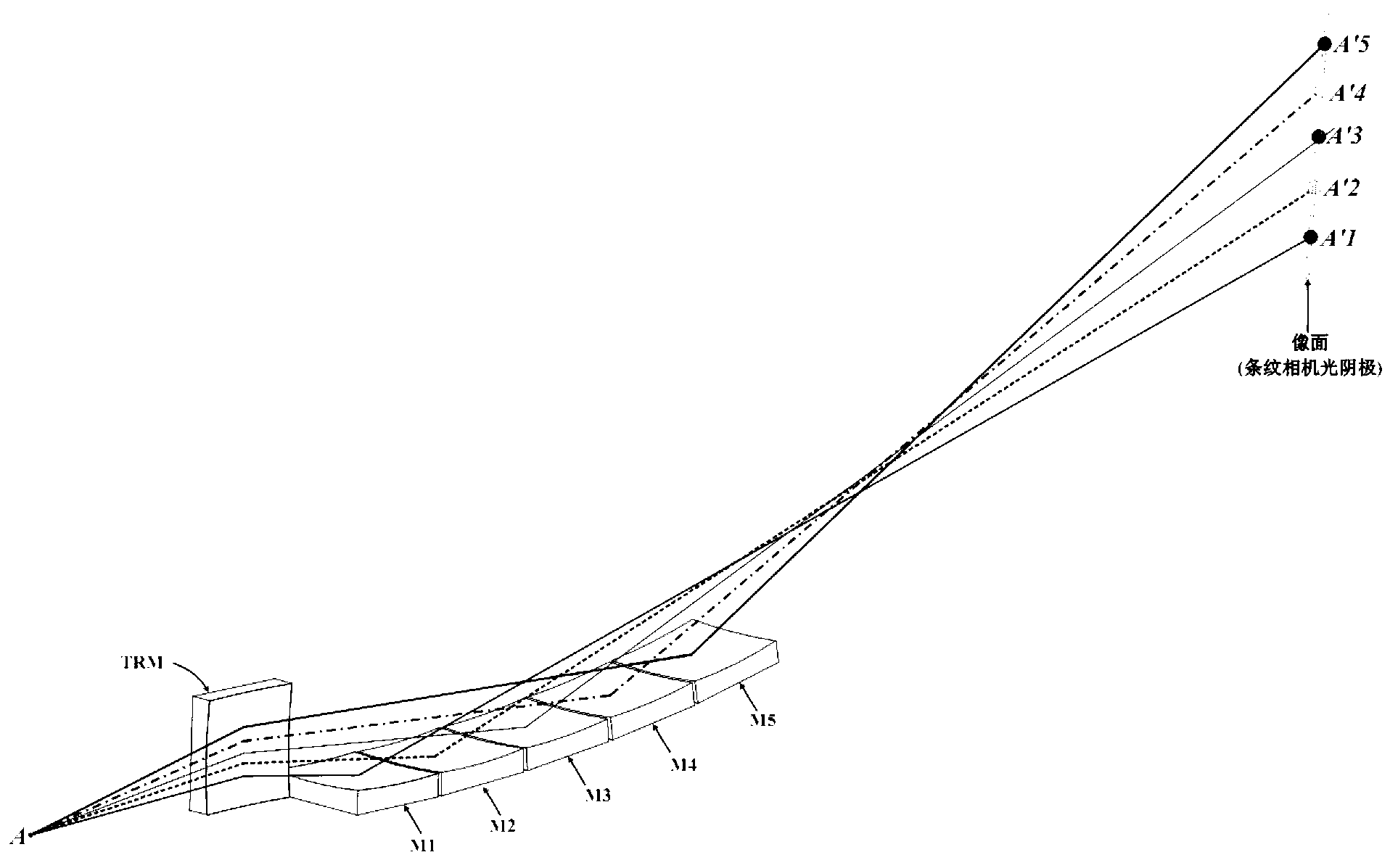
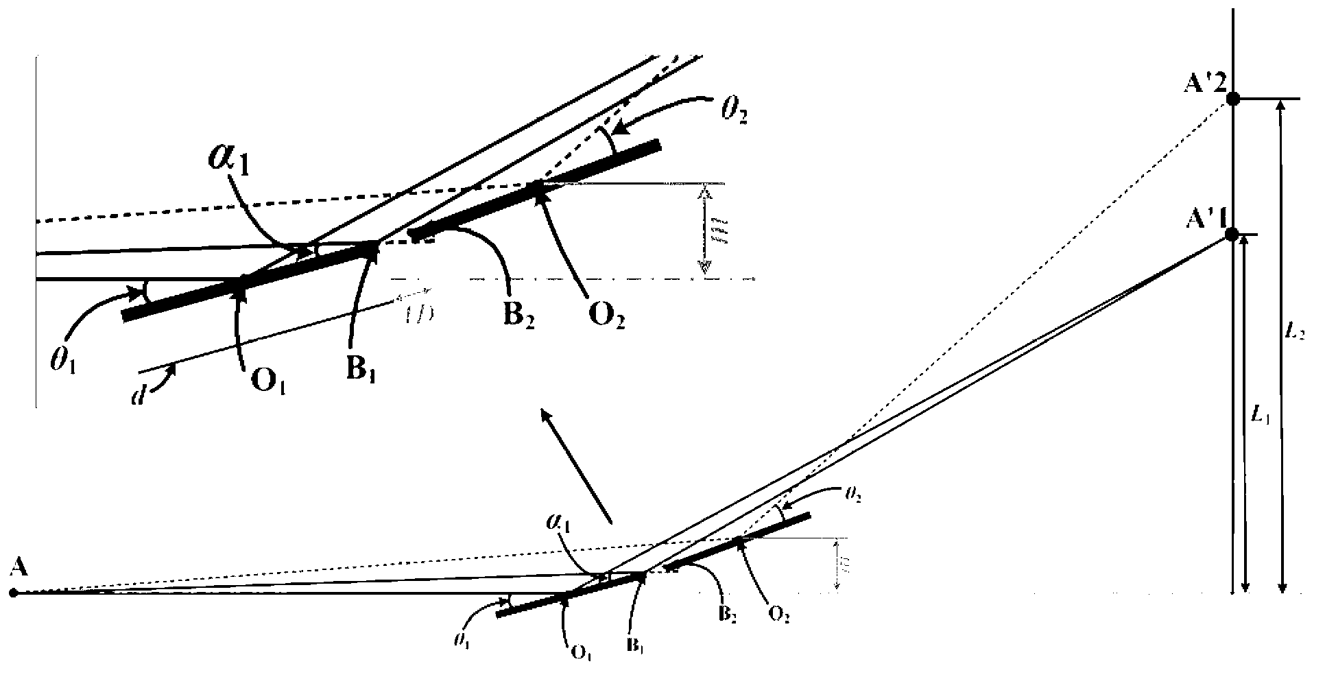
[0020] A time-resolved multicolor monoenergetic X-ray imaging spectrometer, such as figure 1 As shown, the spectrometer includes a spherical objective lens (namely TRM) in the meridional direction and five spherical objective lenses (namely M1, M2, M3, M4, M5) arranged in sequence in the sagittal direction. The spherical objective lenses in the meridional direction are respectively Five channels are composed of five spherical objective lenses in the sagittal direction; the five channels of the spectrometer are all based on the Kirkpatrick-Baez (KB) structure imaging, and the X-rays emitted by the object point are reflected by the spherical objective lens in the meridional direction to form a 3D imaging, and then reflected by 5 spherical objective lenses arranged in sequence in the sagittal direction to form 5-channel 2D imaging on the image plane.

[0021] The spherical objective lens in the meridional direction is coated with a single-layer metal film based on the principle o...

Embodiment 2

[0047] A time-resolved multicolor single-energy X-ray imaging spectrometer, which includes a spherical objective lens in the meridional direction and two spherical objective lenses arranged in sequence in the sagittal direction; the two channels of the spectrometer are based on the Kirkpatrick- Baez (KB) structure imaging, the X-rays emitted by the object point are reflected by the spherical objective lens in the meridional direction to form a one-dimensional imaging, and then reflected by two spherical objective lenses arranged in sequence in the sagittal direction to form two Two-dimensional imaging of the channel.

[0048] The spherical objective lens in the meridional direction is coated with a single-layer metal film based on the principle of total external reflection, and all X-rays with energy lower than the cut-off energy point can be reflected. The spherical objective lens in the sagittal direction is coated with a narrow-band X-ray multilayer film based on the princi...

Embodiment 3

[0051] A time-resolved multicolor single-energy X-ray imaging spectrometer, which includes a spherical objective lens in the meridional direction and 8 spherical objective lenses arranged in sequence in the sagittal direction; the 8 channels of the spectrometer are based on the Kirkpatrick- Baez (KB) structure imaging, the X-rays emitted by the object point are reflected by the spherical objective lens in the meridian direction to form a one-dimensional imaging, and then reflected by 8 spherical objective lenses arranged in sequence in the sagittal direction to form 8 Two-dimensional imaging of the channel.

[0052] The spherical objective lens in the meridional direction is coated with a single-layer metal film based on the principle of total external reflection, and all X-rays with energy lower than the cut-off energy point can be reflected. The spherical objective lens in the sagittal direction is coated with a narrow-band X-ray multilayer film based on the principle of Bra...


PUM


Abstract

The invention relates to a time-resolved multicolor single-energy X-ray imaging spectrometer comprising a spherical objective lens on a meridional direction and n spherical objective lenses sequentially arranged on a sagittal direction, wherein n is an integer larger than 1. n channels are imaged based on a Kirkpatrick-Baez (KB) structure. X-ray emitted by an object point is reflected by the spherical objective lens on the meridional direction, such that a one-dimensional image is formed; and through the reflection of the n spherical objective lenses sequentially arranged on the sagittal direction, a two-dimensional image of n channels is formed on an image plane. Compared with prior art, the multicolor single-energy X-ray imaging spectrometer provided by the invention has higher spatial resolution than existing MMI and bent crystal imaging technology, and higher light collection efficiency than existing MMI and GMXI. Therefore, the spectrometer is suitable for self-luminous imaging diagnosis with low X-ray yield. Especially, the spectrometer can be used on a kJ-level intense laser device at high energy section area or with low energy.

Description

technical field [0001] The invention relates to the field of X-ray imaging, in particular to a time-resolved multicolor single-energy X-ray imaging spectrometer. Background technique [0002] In the research work of laser inertial confinement fusion (ICF), the time-resolved monoenergetic or quasi-monoenergetic imaging observation of laser plasma is an important means to obtain plasma dynamic evolution information, and it is also a necessary way to verify numerical simulation programs. For example, multi-color single-energy X-ray imaging, which uses X-ray single-energy images of multiple energy points in the X-ray energy spectrum range covering the spontaneous luminescence of the target pellet or the characteristic line of a certain doping element, to obtain laser plasma in different regions Important information such as temperature and density distribution. [0003] At present, there are three main multicolor single-energy X-ray imaging methods. The first method is based on...

Claims


Application Information

Patent Timeline
IPC IPC(8): G01N23/04
Inventor 穆宝忠章逸舟伊圣振王新朱京涛王占山
Owner TONGJI UNIV
Why Eureka
Try Eureka