Inlaid digital imaging system of gynecological endoscope

An imaging system and endoscope technology, which is applied in the field of accessories manufacturing, can solve problems such as uneven illumination, difficult image clarity, and large light source attenuation, and achieve the effects of wide observation field, avoiding observation blind spots, and wide field of vision

Inactive Publication Date: 2013-08-14
YANGZHOU SANYUAN TECH
View PDF8 Cites 12 Cited by
  • Summary
  • Abstract
  • Description
  • Claims
  • Application Information

AI Technical Summary

Problems solved by technology

The field of view of the hysteroscope is 60°. Due to the large attenuation of the light source transmitted by the light guide and the unevenness of the illuminated surface, there are certain limitations in eyepiece observation. It is difficult to expand the observation field of view in the uterus and further improve the image clarity.

Method used


Image

  • Inlaid digital imaging system of gynecological endoscope

Examples


Embodiment Construction

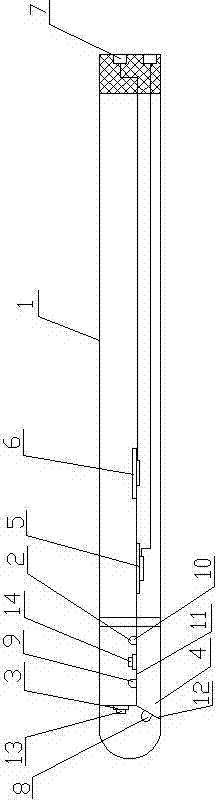
[0012] Such as figure 1 As shown, the gynecological endoscope has a built-in digital imaging system, including an endoscope tube main body 1, an LED light source 2, and a CCD miniature digital camera 3. The front end of the endoscope tube main body 1 is connected to a transparent cover 4, and the LED light source 2. The CCD miniature digital camera 3 is arranged in the transparent cover body 4 respectively, and the power supply module 5 and the digital signal processing module 6 are arranged in the endoscope mirror tube main body 1, and the power supply module 5 is connected with the LED light source 2 and the CCD miniature digital camera respectively. Device 3 is connected, and digital signal processing module 6 is connected with CCD miniature digital camera 3; The LED light source 2 is the first, second and third LED lamp beads 8, 9, 10, and an axial fixed bracket 11 is arranged in the transparent cover body 4, and the second and third LED lamp beads 9, 10 are respective...


PUM


Abstract

The invention relates to a medical instrument, discloses an inlaid digital imaging system of a gynecological endoscope and belongs to the technical field of manufacture of accessories used for electronic digital endoscope medical image workstations of vagina and intrauterine integrated diagnosis and therapy of the gynecological departments. The inlaid digital imaging system of gynecological endoscope comprises an endoscope telescope body, an LED light source and a CCD (charge coupled device) miniature digital camera, the front end of the endoscope telescope body is connected with a transparent cover, the LED light source and the CCD miniature digital camera are respectively arranged in the transparent cover, a power module and a digital signal processing module are arranged in the endoscope telescope body, the power module is respectively connected with the LED light source and the CCD digital camera, the digital signal processing module is connected with the CCD miniature digital camera, a signal output port is arranged at the rear end of the endoscope telescope pipe body, and the signal output port is connected with the digital signal processing module. The inlaid digital imaging system of the gynecological endoscope can be used for examining vagina wall, cervix uteri or even intrauterine and is wide in observation vision field.

Description

technical field [0001] The invention relates to a medical device, in particular to the technical field of manufacturing an accessory of an electronic digital endoscope medical image workstation for integrated diagnosis and treatment of gynecological vagina and uterine cavity. Background technique [0002] In the late 1990s, electronic colposcope was introduced into our country. It is easy to operate and inspect, and has powerful graphic and text functions. It has become a must-have device for gynecological diagnosis and treatment in hospitals at all levels. In its imaging mode, after the speculum expands the inspected surface, through the external light source, the CCD camera can complete the inspection and diagnosis of the cervix and vaginal wall on the exposed field of view of the speculum. Doctors need to configure different types of speculums to meet the requirements of different human body specifications, and the observation site is limited to the cervix. Because the m...

Claims


Application Information

Patent Timeline
Patent Type & Authority Applications(China)
IPC IPC(8): A61B1/05A61B1/303
Inventor 袁传华温汝贤
Owner YANGZHOU SANYUAN TECH
Why Eureka
Try Eureka