
Medical image processing device, method and system based on digital X-ray machine

A medical image and processing device technology, applied in image data processing, medical science, image enhancement, etc., can solve the problems of restricting the application range of CCD image sensors, increasing the burden of data transmission and storage, reducing the accuracy of doctors' diagnosis, and improving the Read and write efficiency, avoid the reduction of storage and processing efficiency, and increase the effect of storage and transmission

Active Publication Date: 2017-07-11
重庆德方信息技术有限公司
View PDF5 Cites 0 Cited by
  • Summary
  • Abstract
  • Description
  • Claims
  • Application Information

AI Technical Summary

Problems solved by technology

[0005] However, when a hospital uses a high-resolution CCD image sensor to take multiple continuous shots of a patient or multiple CCD image sensors to shoot from multiple angles, the amount of collected data is very large. The traditional "image acquisition-PC-terminal control "Device" mode will transmit a large amount of unnecessary information, which increases the burden of data transmission and storage. More importantly, redundant information will greatly interfere with useful information, reducing the accuracy of doctors' diagnosis
[0006] Further, when multiple CCD image sensors are required to work at the same time or take pictures from multiple angles, the multiple transmission lines of traditional wired transmission will seriously restrict the application range of CCD image sensors

Method used


Image

  • Medical image processing device, method and system based on digital X-ray machine
  • Medical image processing device, method and system based on digital X-ray machine
  • Medical image processing device, method and system based on digital X-ray machine

Examples


Embodiment Construction

[0044] In order to make the purpose, technical solution and advantages of the present invention more clear, the present invention will be further described in detail below in conjunction with the accompanying drawings and embodiments.

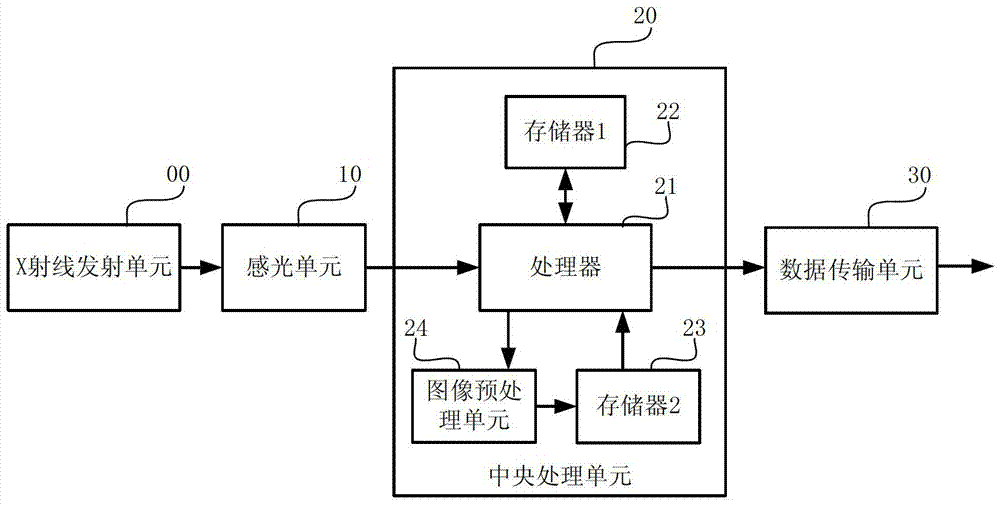
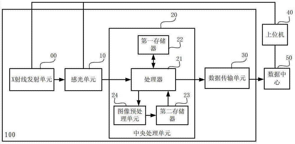
[0045] The present invention provides a medical image processing device 100 for a digital X-ray machine. A schematic diagram of a preferred implementation structure is as follows figure 2 shown, including:

[0046] X-ray emitting unit 00, for emitting X-rays;

[0047] The photosensitive unit 10 is used to convert the X-rays passing through the human body into electrical signals, including a CCD image sensor 11,

[0048] The central processing unit 20 is used to control the original image acquisition, storage and preprocessing of the original image, including a processor 21 and a first memory 22 connected thereto, a second memory 23 and an image preprocessing unit 24; the processor 21 controls the CCD The image sensor 11 collects the original...


PUM


Abstract

The present invention relates to the technical field of medical equipment, and provides a medical image processing device, method and system based on a digital X-ray machine. The device includes an X-ray emitting unit, a photosensitive unit, a central processing unit, and a data transmission unit; the central processing The unit includes a processor and a first memory connected to it, a second memory and an image preprocessing unit; the first memory and the second memory are separately set, one is used to store the original image information, and the other is used to store the preprocessed image After the image information, the access of the original image is separated from the writing and reading of the preprocessed image, which improves the efficiency of medical image data reading and writing; the image preprocessing unit preprocesses the original image, including the original image. Compression processing, noise reduction processing, and effective gray level selection processing eliminate redundant information in the original image, increase the storage and transmission of useful information, and improve transmission efficiency.

Description

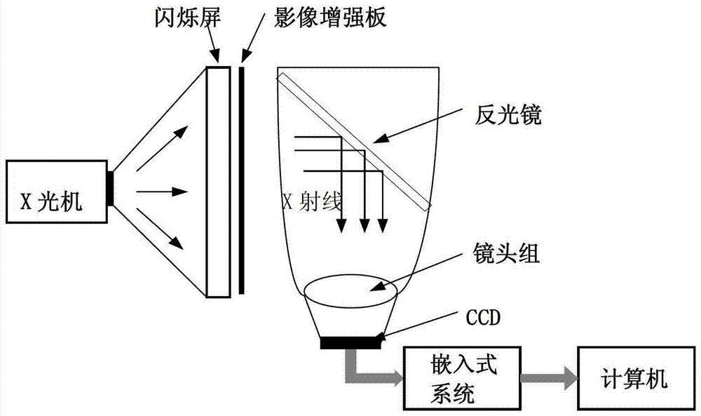
technical field [0001] The invention relates to the technical field of medical devices, in particular to a medical image processing device, method and system based on a digital X-ray machine. Background technique [0002] In modern medicine, medical images are important data for doctors to judge a patient's condition and are crucial to diagnosis. Now there are some known methods for medical image acquisition and processing. [0003] Medical image acquisition is usually as figure 1 The schematic diagram of the structure shown in the figure shows that due to the invisible characteristics of X-rays, the charge-coupled device CCD camera cannot directly collect X-rays, so the X-rays are converted into weak visible light through the scintillation screen, and the weak light signal is enhanced by the image intensifier board to convert The final light is visible light, and the visible light is collected by the large acceptance and short focal length lens group to the CCD camera afte...

Claims


Application Information

Patent Timeline
Patent Type & Authority Patents(China)
IPC IPC(8): G06T5/00A61B6/00
Inventor 李章勇刘圣蓉刘杰张荔芳王伟赵志强赵德春庞宇王浩
Owner 重庆德方信息技术有限公司
Learn More