

Image force microscopy of molecular resonance
An atomic force microscope, molecular technology, applied in scanning probe microscopy, manipulation of single atoms, instruments, etc., can solve the problem of spectral identification without the use of detection materials
- Summary
- Abstract
- Description
- Claims
- Application Information
AI Technical Summary
Problems solved by technology
Method used
Image

Examples
Embodiment Construction
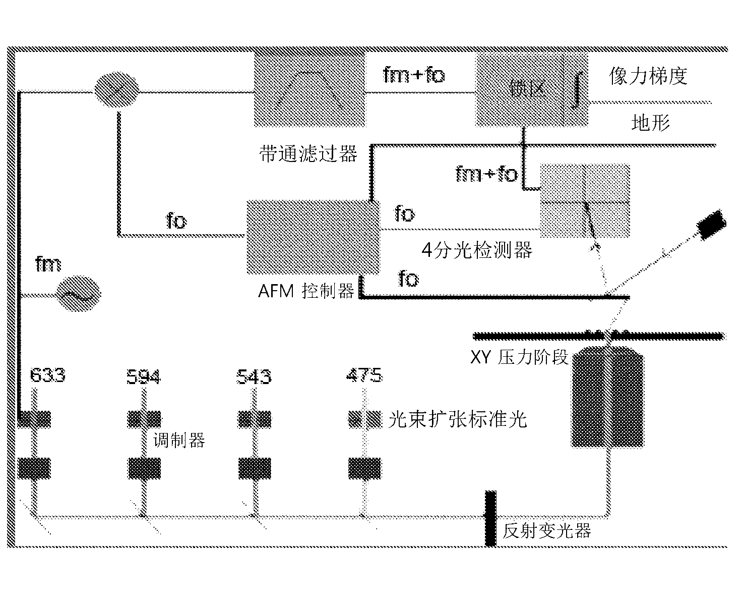
[0026] The cantilever provides the 1st mechanical resonance f 0 ( figure 1 ) in the gravitational mode (attractive mode) AFM of vibration 7 microscope. f 0 Changes in the oscillation amplitude detect the van der Waal (van der Waal) gravitational force between the tip and the object, and can also use and obtain topography feedback signals. In the traditional tapping (tapping) mode AFM, the AFM detector is firmly on the object. Using a stiffness constant with k=3n / m and f 0 = 65KHz of the 1st mechanical resonance of the cantilever. choose f m f = 360KHz laser modulation frequency is detected 0 +f m The frequency in the upper sideband is 425KHz.
[0027] Different from traditional methods, in changing the frequency f of the force gradient between object features m Excitation / laser beam (energy source) in and changing f m mirror image in . This force gradient modulation follows from f 0 +f m and f 0 -f m Frequency-sequential modulation of cantilever mechanical reso...
PUM
 Abstract
Description
Claims
Application Information