Method for measuring depth and three-dimensional size of nidus tissue by capsule endoscope

A technology of capsule endoscopy and measurement methods, which can be used in diagnostic recording/measurement, medical science, sensors, etc., and can solve problems such as long calculation time, unsuitable long time, and need to improve accuracy and speed

Inactive Publication Date: 2013-09-18
NINGBO INST OF TECH ZHEJIANG UNIV ZHEJIANG
View PDF4 Cites 21 Cited by
  • Summary
  • Abstract
  • Description
  • Claims
  • Application Information

AI Technical Summary

Problems solved by technology

The advantage of this positioning technology is that it utilizes the existing radio frequency signal and antenna in the wireless capsule endoscope, without adding any components in the wireless capsule endoscope; however, the positioning accuracy of this positioning technology is very low, and the average positioning accuracy is 37.7mm, unacceptable for clinical application
Medical imaging technologies such as X-ray imaging, CT and MRI (Magnetic Resonance Imaging, Magnetic Resonance Imaging) can also use 3D reconstruction technology to locate, but the process is complicated and the calculation time is long; and the position or direction is obtained by indirect reconstruction calculation technology, Accuracy and speed are limited; additionally there are radiation damage and time-consuming issues
3D ultrasonic image technology can also be used for positioning, which uses a tracking device

Method used


Image

  • Method for measuring depth and three-dimensional size of nidus tissue by capsule endoscope
  • Method for measuring depth and three-dimensional size of nidus tissue by capsule endoscope
  • Method for measuring depth and three-dimensional size of nidus tissue by capsule endoscope

Examples


Embodiment 1

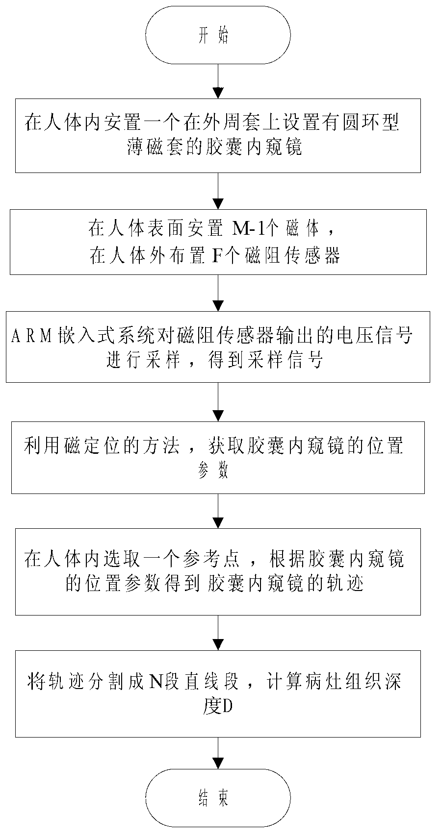
[0100] In this embodiment, a method for measuring the tissue depth of a capsule endoscope lesion is proposed, and the flow chart of the method is as follows figure 1 shown, including the following steps:

[0101] ① Obtain the accurate position parameters of the capsule endoscope by placing magnets and magnetoresistive sensors. The specific process is as follows:

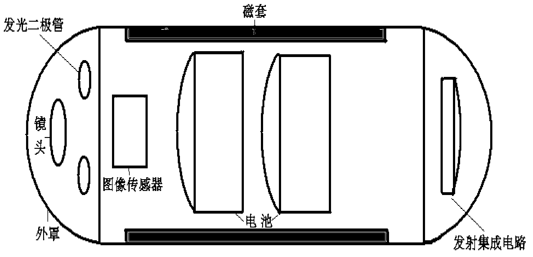
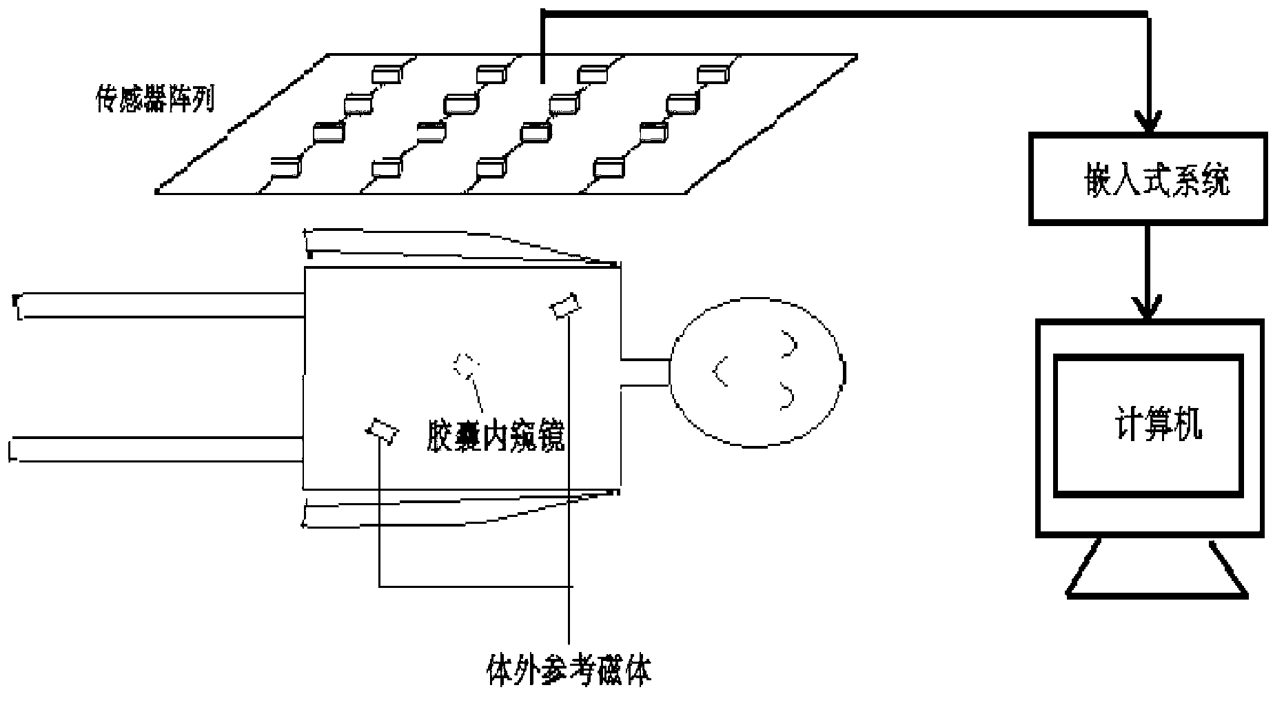
[0102] ①-1, such as image 3 , establish a space coordinate system outside the human body as a reference coordinate system, place a capsule endoscope with a ring-shaped thin magnetic sleeve on the outer peripheral sleeve inside the human body, the structure of the capsule endoscope is as follows figure 2 As shown, before obtaining the position parameters of the capsule endoscope, M-1 magnets are placed on the surface of the human body, together with the annular thin magnetic sleeve of the capsule endoscope to form M magnets, which are determined by the arrangement of M magnets The three-dimensional position of the...

Embodiment 2

[0125] In this embodiment, a method for measuring the three-dimensional size of the lesion tissue under capsule endoscopy is proposed, and its flow chart is as follows Figure 7 shown, including the following steps:

[0126] (1) Accurate position parameters and direction parameters of the capsule endoscope before and after the movement of the capsule endoscope and the rotation of the capsule endoscope around the central axis after the movement of the capsule endoscope are acquired by placing magnets and magnetoresistive sensors Angle, the specific process is as follows:

[0127] ⑴-1, such as image 3 , establish a spatial coordinate system outside the human body as a reference coordinate system, place an image sensor outside the human body, and place a capsule endoscope with a ring-shaped thin magnetic sleeve on the outer peripheral sleeve inside the human body. The capsule endoscope structured as figure 2 As shown, before obtaining the position parameters of the capsule e...


PUM


Abstract

The invention provides a method for measuring depth and three-dimensional size of a nidus tissue by a capsule endoscope, which comprises the following steps: a location parameter of the capsule endoscope is obtained by utilizing a magnetic positioning method, then corresponding to a start point, a track of the capsule endoscope is obtained according to the location parameter of the capsule, the track is divided to various sections of straight lines, and the depth, corresponding to start point, of the nidus tissue is ensured by calculating each straight line; and images photographed before and after motions of the capsule endoscope is utilized to obtain a rotation angle of the capsule endoscope around a central main shaft after motion of the capsule endoscope, a three-dimensional location of feature points in the nidus tissue is calculated by the location parameter and a direction parameter of the capsule endoscope obtained by combining with the magnetic localization method, and the three-dimensional size of the nidus tissue is obtained according to the three-dimensional location of the feature points. The method has the advantages of being capable of weakening or eliminating the influence of human motion for the working of the capsule endoscope in a human body, and accurately realizing the measuring for the depth and the three-dimensional size of the nidus tissue.

Description

technical field [0001] The invention relates to a capsule endoscope technology, in particular to a method for measuring the depth and three-dimensional size of a capsule endoscope lesion tissue. Background technique [0002] Israel's Given Imaging company proposed a positioning technology based on wireless radio frequency RF signals, which uses 8 antennas outside the human body to receive radio frequency RF signals from the wireless capsule endoscope, and uses algorithms to calculate the position of the wireless capsule endoscope . The advantage of this positioning technology is that it utilizes the existing radio frequency signal and antenna in the wireless capsule endoscope, without adding any components in the wireless capsule endoscope; however, the positioning accuracy of this positioning technology is very low, and the average positioning accuracy is 37.7mm, unacceptable for clinical application. Medical imaging technologies such as X-ray imaging, CT and MRI (Magneti...

Claims


Application Information

Patent Timeline
IPC IPC(8): A61B5/107A61B5/06
Inventor 胡超苗厚想何小其冯忠晴袁小英陈建可王永辉
Owner NINGBO INST OF TECH ZHEJIANG UNIV ZHEJIANG
Why Eureka
Try Eureka