Flow type fluorescence microscopy imaging device and method

A microscopic imaging and flow fluorescence technology, which is applied in the direction of measuring devices, fluorescence/phosphorescence, instruments, etc., can solve the problems of affecting the real-time performance of the system, imaging tailing of moving objects, and taking up time, etc.

Inactive Publication Date: 2013-09-18
香港浸会大学深圳研究院
View PDF0 Cites 19 Cited by
  • Summary
  • Abstract
  • Description
  • Claims
  • Application Information

AI Technical Summary

Problems solved by technology

However, the deconvolution operation will take time and affect the real-time performance of the system.
[0006] The second major problem is tailing from flowing samples
[0008] The present invention proposes a new flow type fluorescent microscopic imaging device and imaging method, which can well solve the problem of small focal depth and tailing problems when imaging moving objects in the existing imaging flow cytometer

Method used


Image

  • Flow type fluorescence microscopy imaging device and method
  • Flow type fluorescence microscopy imaging device and method
  • Flow type fluorescence microscopy imaging device and method

Examples


Embodiment Construction

[0025] The present invention will be further described below in conjunction with the accompanying drawings and embodiments.

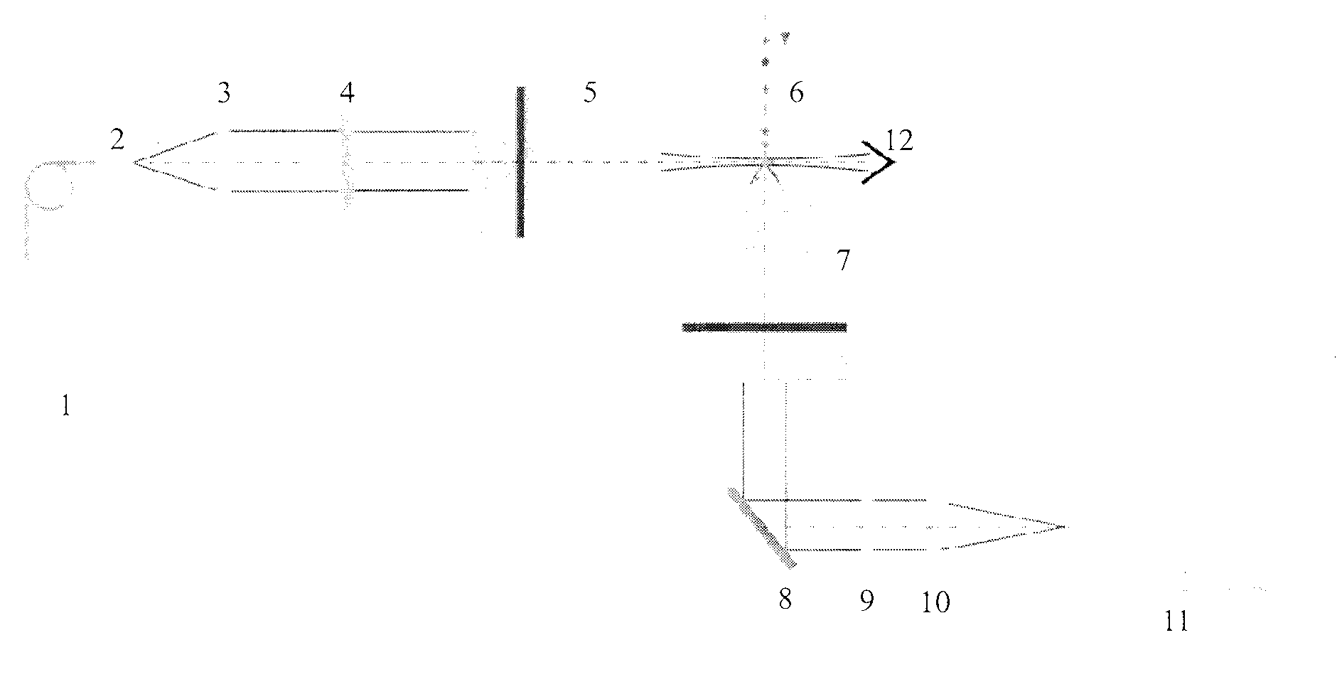
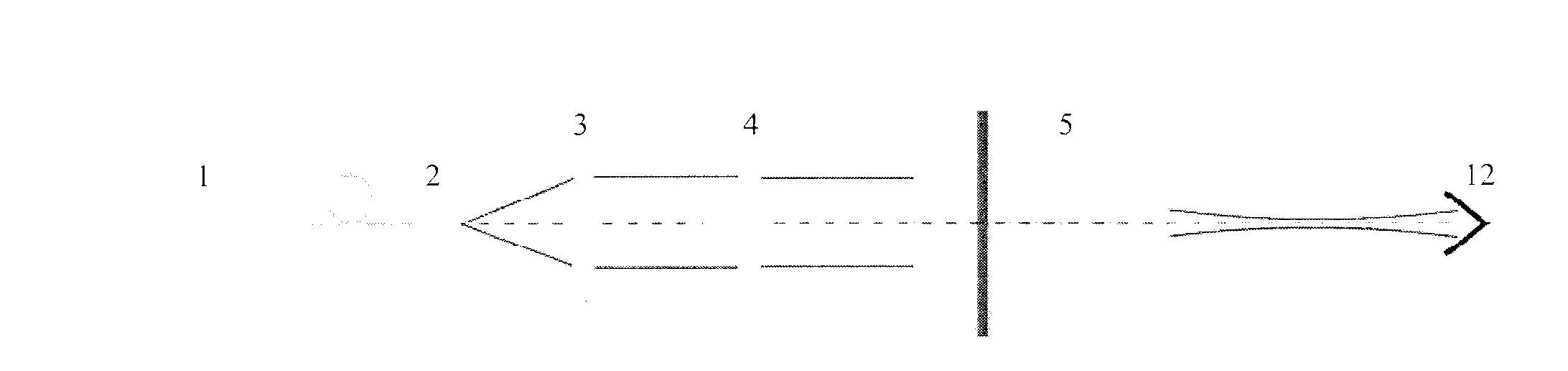
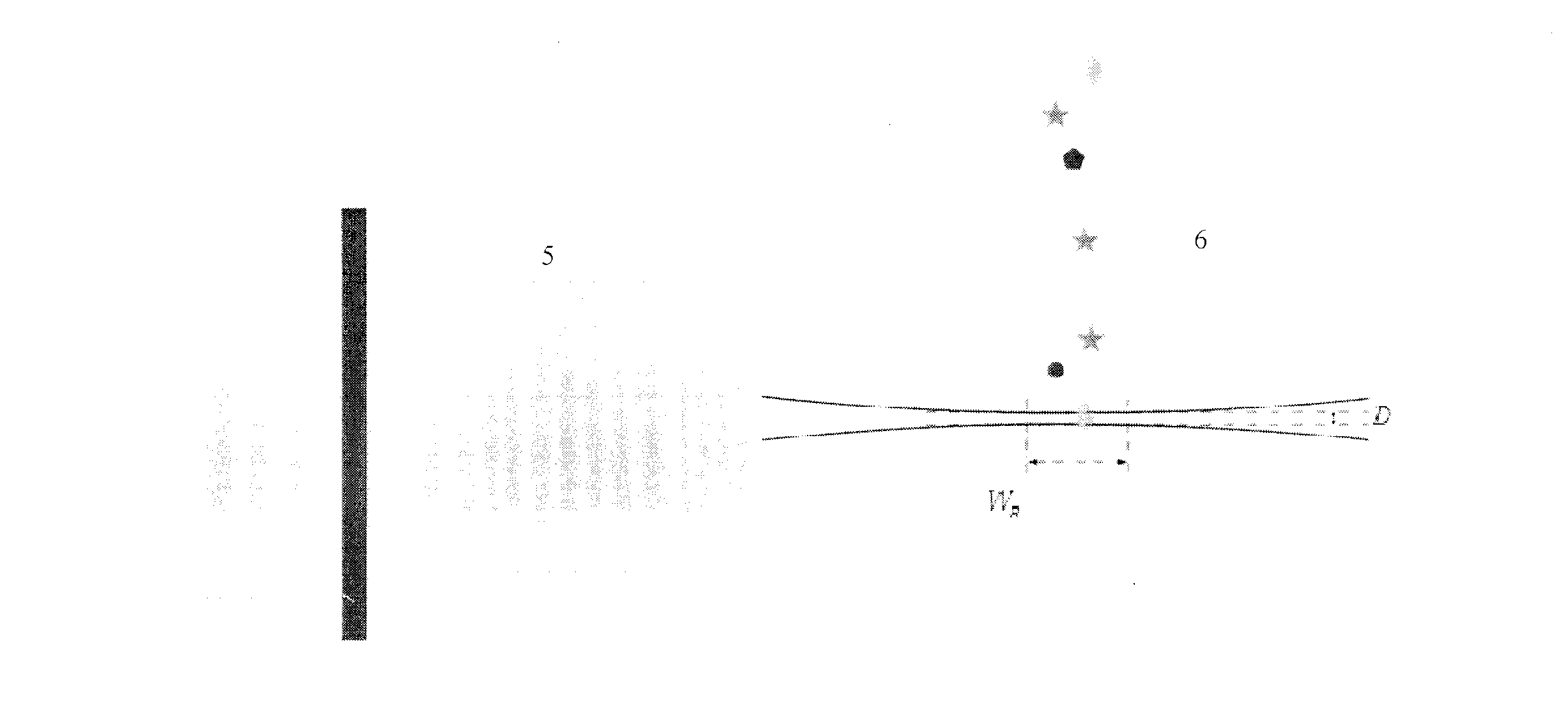
[0026] The invention is a flow type fluorescent microscopic imaging device, which can solve the problems of small focus depth of imaging flow cytometer and trailing when imaging moving objects. The beneficial effects of the present invention include: 1) using a sheet light source with a thickness close to the diffraction limit to excite the autofluorescence or non-autofluorescence of cells can overcome the problem of small depth of focus in fluorescence microscopy imaging; The cells are excited and emit fluorescence, which can effectively reduce the background of the fluorescence image, so it can effectively improve the sensitivity of the system; 3) Cells pass through the excitation light source vertically, and the imaging direction is parallel to the sample flow direction to perform fluorescence imaging on the cells, which can The tailing phenomenon is...


PUM


Abstract

The invention discloses a flow type fluorescence microscopy imaging device, in order to solve the problems of small focal depth of an imaging flow cytometer as well as trailing which occurs when a moving object is imaged. The imaging device has the following beneficial effects that (1) auto-fluorescence or non-auto-fluorescence of cells is excited by a light sheet source with the thickness near a diffraction limit, so that the problem of small focal depth of fluorescence microscopy imaging can be solved; (2) only the cells positioned within the range of focal depth are excited to emit fluorescence and a background of a fluorescence image can be lowered effectively, so that the sensitivity of a system can be enhanced effectively; (3) the cells perpendicularly pass through an excitation light source and are subjected to fluorescence imaging in a way that the imaging direction is parallel to the flowing direction of a sample, so that the trailing phenomenon can be well inhibited; (4) multiple cells are allowed to be detected simultaneously, so that the detection speed of the system can be increased.

Description

technical field [0001] The present application relates to a flow fluorescence microscopy imaging device, more specifically to a fluorescence microscopy imaging device combining single-layer light-excited fluorescence microscopy and flow cytometry. Background technique [0002] In the fields of biology and medicine, flow cytometry is the main detection tool when large numbers of cells need to be scanned. Traditional flow cytometers observe cells as points, and can obtain high detection speed without completely sacrificing spatial resolution. Currently, its detection speed can reach tens of thousands of cells per second. Imaging flow cytometry can not only obtain the "point" information obtained by traditional flow cytometry, but also perform microscopic imaging of cells to obtain spatial information of cells. Compared with traditional flow cytometers, imaging flow cytometers have greater advantages when it is necessary to obtain information about the morphology and internal...

Claims


Application Information

Patent Timeline
Patent Type & Authority Applications(China)
IPC IPC(8): G01N15/14G01N21/64
CPCG01N2015/144G01N15/1434
Inventor 吴江来陈启尧李剑平
Owner 香港浸会大学深圳研究院
Why Eureka
Try Eureka