A method and processing device for suppressing bone images in X-ray images
A line image and image technology, applied in the field of suppressing bone images in X-ray images, methods and processing equipment, can solve the problem that the single-pixel prediction model cannot effectively describe the pixel correlation, it is difficult to ensure the spatial consistency of generated soft tissue images, and it is difficult to effectively use Problems such as the global information of the chest radiograph image, to achieve the effects of bone image suppression, image quality index optimization, and improved effect
- Summary
- Abstract
- Description
- Claims
- Application Information
AI Technical Summary
Problems solved by technology
Method used
Image
Examples
Embodiment 1
[0044]The invention provides a method for suppressing bone images in X-ray images without performing dual-energy subtraction photography. Specifically, the present invention uses the previous DES image data to predict and reconstruct the bone image for a single X-ray image taken by ordinary DR or CR equipment, and generates a soft tissue image through subtraction; without increasing the exposure dose and using DES equipment , to achieve the suppression of bone images in X-ray images.
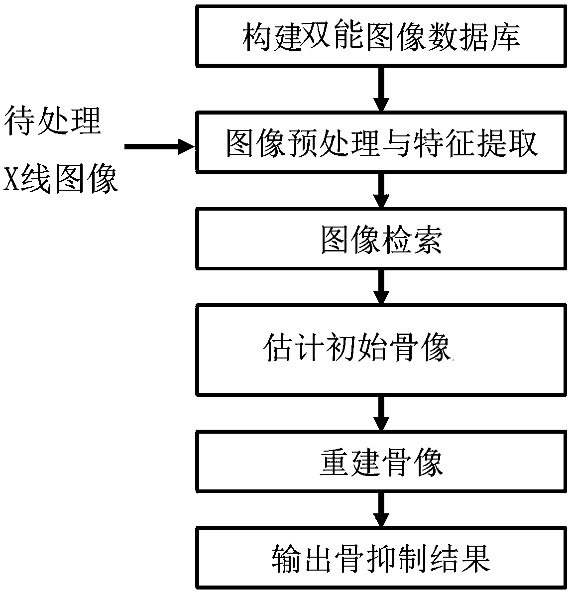
[0045] A method of suppressing bone images in X-ray images of the present invention, such as figure 1 As shown, the following steps are sequentially included.
[0046] (1) Preprocess the X-ray image to be processed and extract its image features, and the extracted image features are called subject image features.
[0047] Among them, the preprocessing of the X-ray image to be processed is specifically to perform uniform and standardized processing on the gray value range, spatial resolution, a...
Embodiment 2
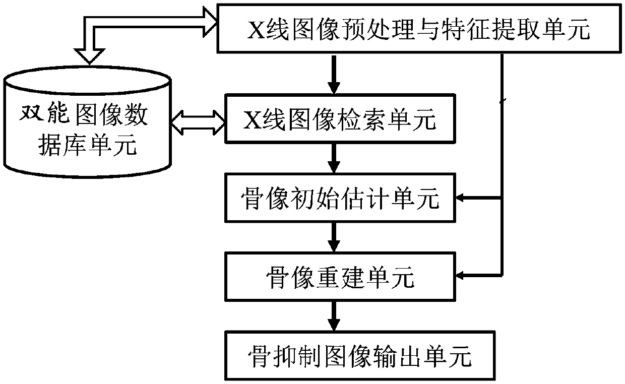
[0060] A processing device for suppressing skeletal images in x-ray images, such as figure 2 As shown, the method for suppressing bone images in the X-ray image of the above-mentioned embodiment 1 is used for image processing, and the processing unit provided includes: a dual-energy image database unit, an X-ray image preprocessing and feature extraction unit, an X-ray image retrieval unit, A bone image initial estimation unit, a bone image reconstruction unit and a bone suppression image output unit.
[0061] The dual-energy image database unit pre-stores a number of clinical dual-energy subtraction prior reference X-ray images and corresponding soft tissue images, bone images, lesion area location information and image feature information of the prior reference X-ray images. The information stored in the dual-energy image database unit can be stored in advance, and can also be imported when used.
[0062] The X-ray image preprocessing and feature extraction unit preprocess...
Embodiment 3
[0069] The method of the present invention is described with a specific embodiment.
[0070] Pre-preparation is done in advance, and the dual-energy image database unit is pre-stored with clinically real DES image data, including image information of conventional chest X-rays and their corresponding soft tissue and bone images, imaging parameters, nodules and lesion size and location data, etc. information, and these data information are stored in the memory of the dual-energy image database unit.
[0071] To process the X-ray image information to be processed, the specific steps are as follows.
[0072] (1) Preprocess the X-ray image to be processed and extract its image features, and the extracted image features are called subject image features. The subject characteristics may be stored in the memory of the dual-energy image database unit.
[0073] First, the gray value range, spatial resolution, and contrast of the X-ray images to be processed are processed according to ...
PUM
 Abstract
Description
Claims
Application Information
 - R&D
- Intellectual Property
- Life Sciences
- Materials
- Tech Scout
- Unparalleled Data Quality
- Higher Quality Content
- 60% Fewer Hallucinations
Browse by: Latest US Patents, China's latest patents, Technical Efficacy Thesaurus, Application Domain, Technology Topic, Popular Technical Reports.
© 2025 PatSnap. All rights reserved.Legal|Privacy policy|Modern Slavery Act Transparency Statement|Sitemap|About US| Contact US: help@patsnap.com


