
Double-lens-cone microscope device used in urinary sediment inspection equipment

A detection equipment and double-lens tube technology, applied in microscopes, optics, instruments, etc., can solve problems such as high assembly accuracy, high processing accuracy requirements for objective lens mounting holes, and misalignment, so as to reduce assembly requirements and production costs. Save the time of taking pictures and improve the detection speed

Active Publication Date: 2013-11-27
URIT MEDICAL ELECTRONICS CO LTD
View PDF7 Cites 8 Cited by
  • Summary
  • Abstract
  • Description
  • Claims
  • Application Information

AI Technical Summary

Problems solved by technology

[0003] The traditional microscope uses the card or ball inside the objective lens converter to realize the positioning of the objective lens during the high-low magnification conversion process, but this way of realizing the high-low magnification conversion by the objective lens converter has the following shortcomings: (1) The installation of the objective lens of the objective lens converter The hole machining accuracy is required to be high, otherwise it is difficult to ensure the coaxiality between the objective lens and the lens barrel, especially when the magnification is large; (2) When the motor and gear transmission are added to control the objective lens converter to automatically realize the high and low magnification conversion, The assembly accuracy is required to be high, otherwise it is easy to have return errors and misalignment
In addition, in traditional microscopes, the light source (halogen lamp and LED lamp) is directly placed under the counting cell, so heat will inevitably be generated, which will have an unpredictable impact on the environment of the entire microscope and the detection results, especially the image at high magnification

Method used


Image

  • Double-lens-cone microscope device used in urinary sediment inspection equipment
  • Double-lens-cone microscope device used in urinary sediment inspection equipment
  • Double-lens-cone microscope device used in urinary sediment inspection equipment

Examples


Embodiment Construction

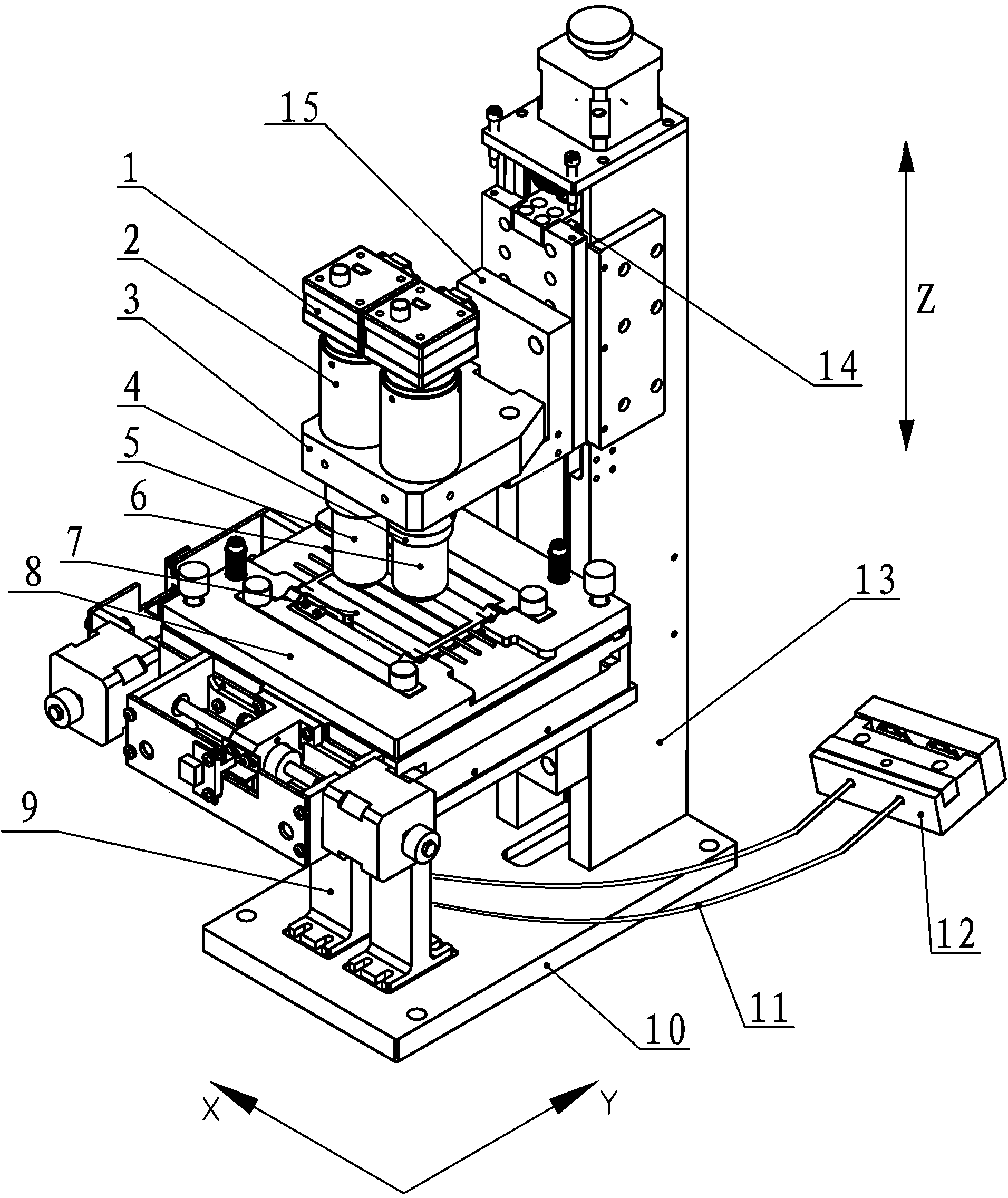
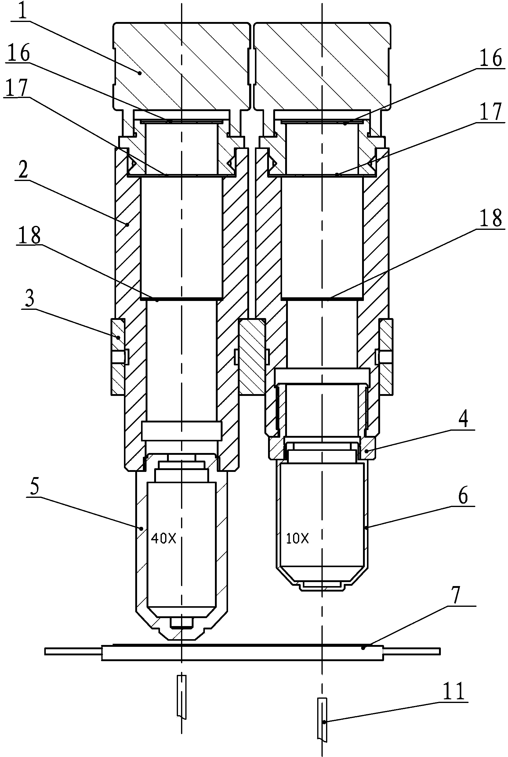
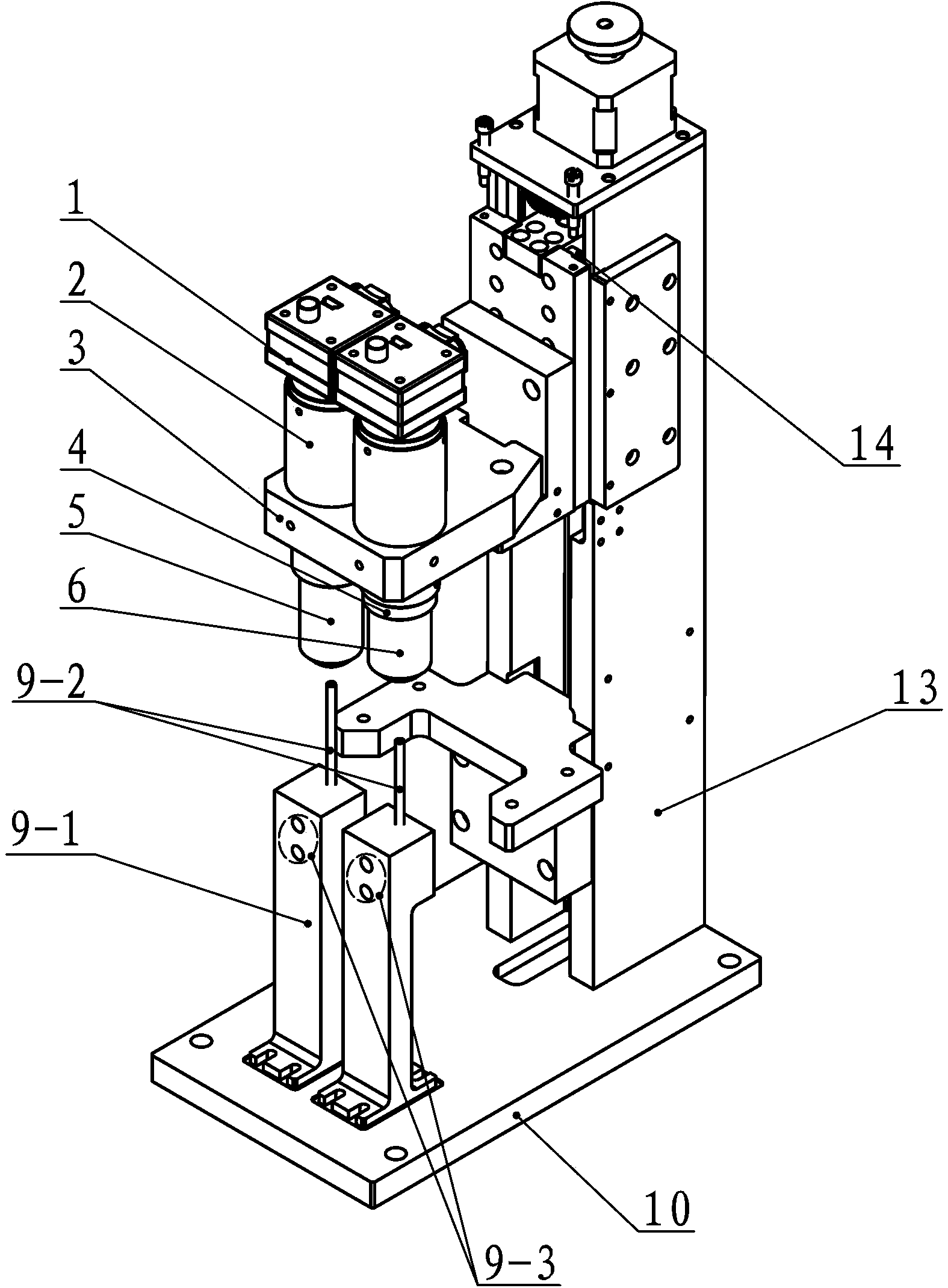
[0024] Such as figure 1 As shown, the double-lens tube microscope device used in urine sediment detection equipment according to the present invention includes a support block 3, a high-magnification objective lens 5, a low-magnification objective lens 6, a counting pool 7 and a light source, and also includes a support block 3 2 objective lens connecting tubes 2 on the top, and 2 CCD sensors 1 respectively installed above the objective lens connecting tube 2; where:

[0025] The support block 3 is installed on a guide rail 13 of a column 12 with a base 10 through a slide seat 15, and the support block 3 can reciprocate in the longitudinal direction through the cooperation of the slide block and the guide rail 13;

[0026] The counting pool 7 is arranged on the stage 8 (the stage 8 is preferably able to move in the horizontal direction (X-axis direction and Y-axis direction), and its structure is referred to the utility model patent whose publication number is CN202837670U ),...


PUM

PropertyMeasurementUnit
Heightaaaaaaaaaa

Abstract

The invention discloses a double-lens-cone microscope device used in urinary sediment inspection equipment. The double-lens-cone microscope device comprises a supporting block, a high power objective, a low power objective, a counting cell, a light source, two objective connecting cylinders and two CCD sensors, wherein the two objective connecting cylinders are installed on the supporting block, and the two CCD sensors are installed above the objective connecting cylinders respectively. Three diaphragms are arranged inside each objective connecting cylinder at intervals. The light source is placed at the position far away from the counting cell, the light source comprises two LED light source bodies and two optical fibers, one end of each optical fiber is located below the counting cell and directly faces the counting cell, and the other end of each optical fiber directly faces the corresponding LED light source body. The parts form a high power microscope system and a low power microscope system respectively, and the central axes of the CCD sensor, the objective connecting cylinder, the high power objective or the low power objective and the light source optical fiber which all belong to the same set of microscope system coincide. The double-lens-cone microscope device is simple in structure, can shoot visible components in urine under different powers at the same time, saves shooting time and improves inspection speed.

Description

technical field [0001] The invention relates to the technical field of image processing of a microscope system, in particular to a double lens barrel microscope device used in urine sediment detection equipment. Background technique [0002] In the detection equipment of urine formed components, traditional microscopes use a lens barrel containing multiple sets of optical lenses, and use a porous objective lens converter to realize manual or automatic conversion between objective lenses of various magnifications or use a zoom lens to change the imaging magnification , so as to obtain the images of the detected object under different magnifications; the light sources used are generally halogen lamps and LED lights directly injected into the objective lens. [0003] The traditional microscope uses the card or ball inside the objective lens converter to realize the positioning of the objective lens during the high-low magnification conversion process, but this way of realizing ...

Claims


Application Information

Patent Timeline
IPC IPC(8): G02B21/24G02B21/36G02B21/06G01N15/04
Inventor 郑彬陈祥云唐雪辉农柳华蒋均黄联庆
Owner URIT MEDICAL ELECTRONICS CO LTD
Learn More