Ultrasonic diagnostic apparatus
A diagnostic device, ultrasonic technology, applied in the direction of acoustic wave diagnosis, infrasonic wave diagnosis, ultrasonic/sonic wave/infrasonic wave diagnosis, etc., can solve problems such as deterioration and reduction, and achieve the effect of optimizing image quality
- Summary
- Abstract
- Description
- Claims
- Application Information
AI Technical Summary
Problems solved by technology
Method used
Image

Examples
no. 1 Embodiment approach )
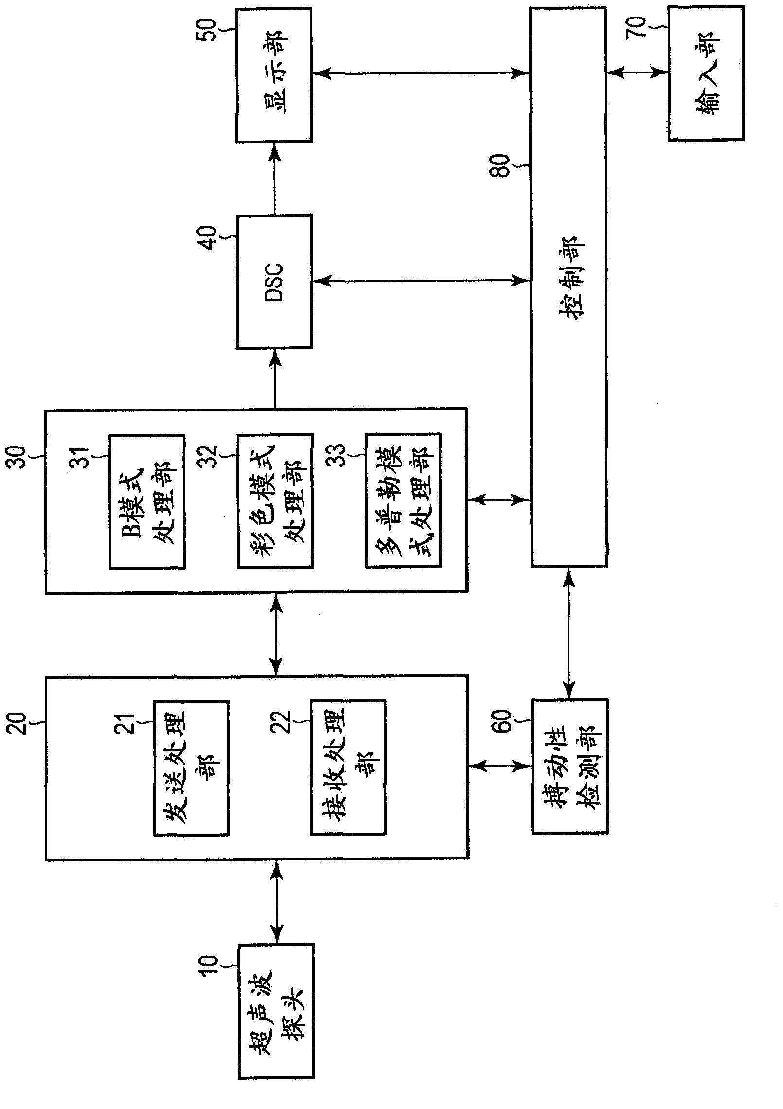
[0036] First, the ultrasonic diagnostic apparatus according to the first embodiment will be described. figure 1 The configuration of the ultrasonic diagnostic apparatus according to this embodiment is shown.
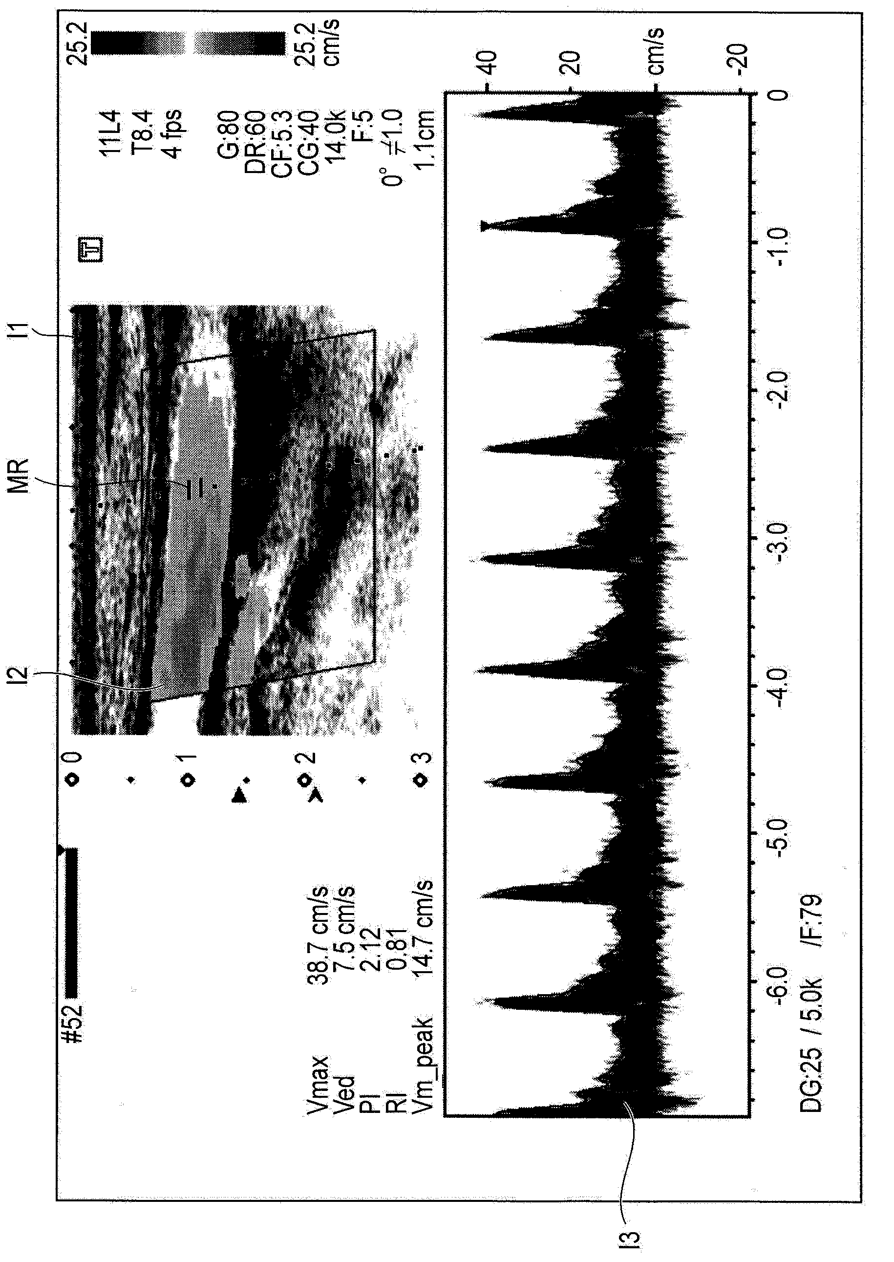
[0037] The ultrasonic diagnostic apparatus according to this embodiment has a function of simultaneously displaying images of a plurality of operation modes including the Doppler mode, such as the triple mode. The ultrasonic diagnostic apparatus according to this embodiment will be described below as a plurality of operating modes that simultaneously display images in the B-mode, color mode, and Doppler mode. The B-mode is a mode for displaying a two-dimensional morphological image (B-mode image). The color mode is a mode for displaying a two-dimensional blood flow image (color Doppler mode image). In addition, the Doppler mode is a mode for displaying a Doppler waveform (Doppler spectral image).
[0038] In addition, in the ultrasonic diagnostic apparatus of the pres...
no. 2 Embodiment approach )
[0082] Next, an ultrasonic diagnostic apparatus according to a second embodiment will be described. Similar to the first embodiment described above, the ultrasonic diagnostic apparatus according to this embodiment has been described as displaying images of the B-mode, the color mode, and the Doppler mode simultaneously as a plurality of operation modes including the Doppler mode.
[0083] In addition, since the configuration of the ultrasonic diagnostic apparatus according to this embodiment is the same as that of the above-mentioned first embodiment, it is appropriate to use figure 1 Be explained. Here, the description will mainly focus on the parts that are different from the above-mentioned first embodiment.
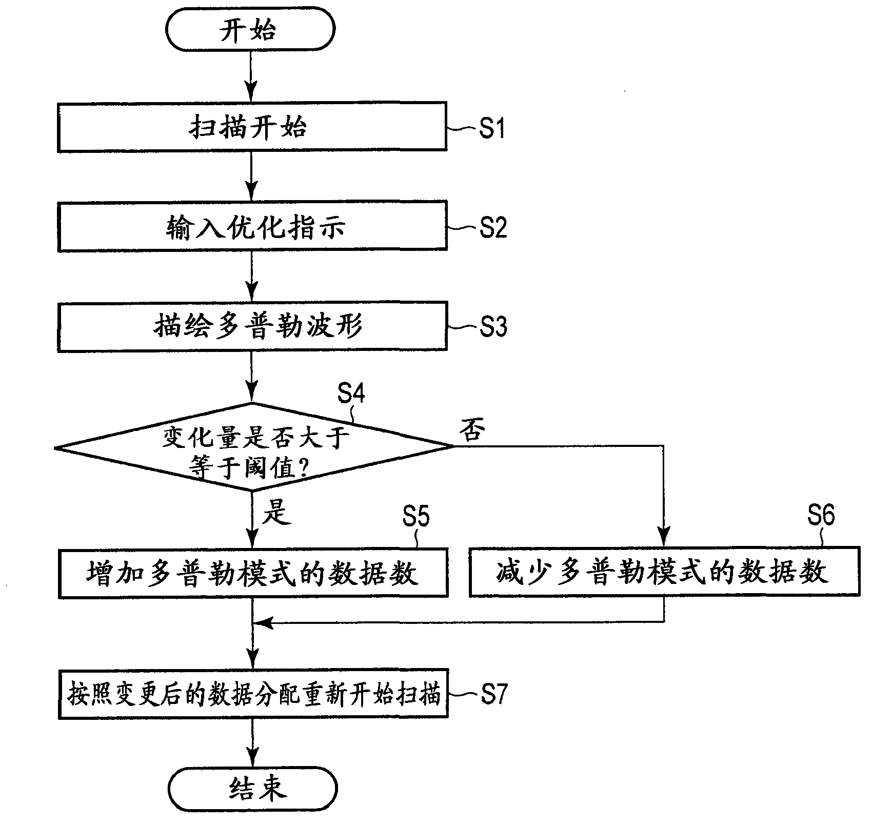
[0084] In the above-mentioned first embodiment, an example was described in which the data distribution of each operation mode is changed according to the determination result of whether the subject's blood flow is a pulsating flow or a steady flow, but in this embod...
PUM
 Abstract
Description
Claims
Application Information
 - R&D
- Intellectual Property
- Life Sciences
- Materials
- Tech Scout
- Unparalleled Data Quality
- Higher Quality Content
- 60% Fewer Hallucinations
Browse by: Latest US Patents, China's latest patents, Technical Efficacy Thesaurus, Application Domain, Technology Topic, Popular Technical Reports.
© 2025 PatSnap. All rights reserved.Legal|Privacy policy|Modern Slavery Act Transparency Statement|Sitemap|About US| Contact US: help@patsnap.com