Endoscope device and measuring method
An endoscope and measuring point technology, applied to endoscopes, measuring devices, optical devices, etc., can solve problems such as the accuracy of measurement results and the decrease in inspection efficiency, and achieve the effect of easy confirmation
- Summary
- Abstract
- Description
- Claims
- Application Information
AI Technical Summary
Problems solved by technology
Method used
Image
Examples
no. 1 approach
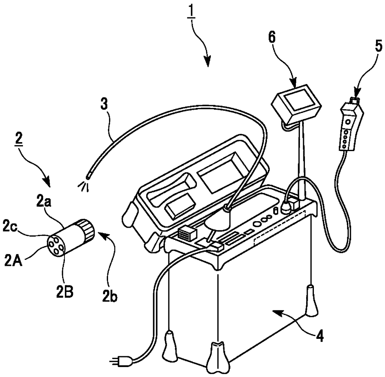
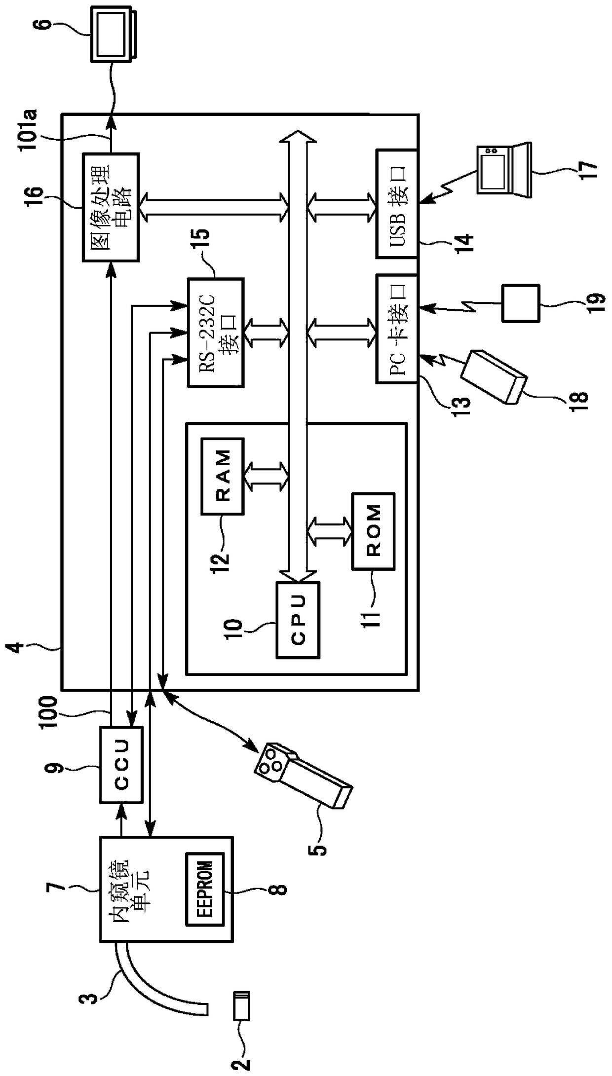
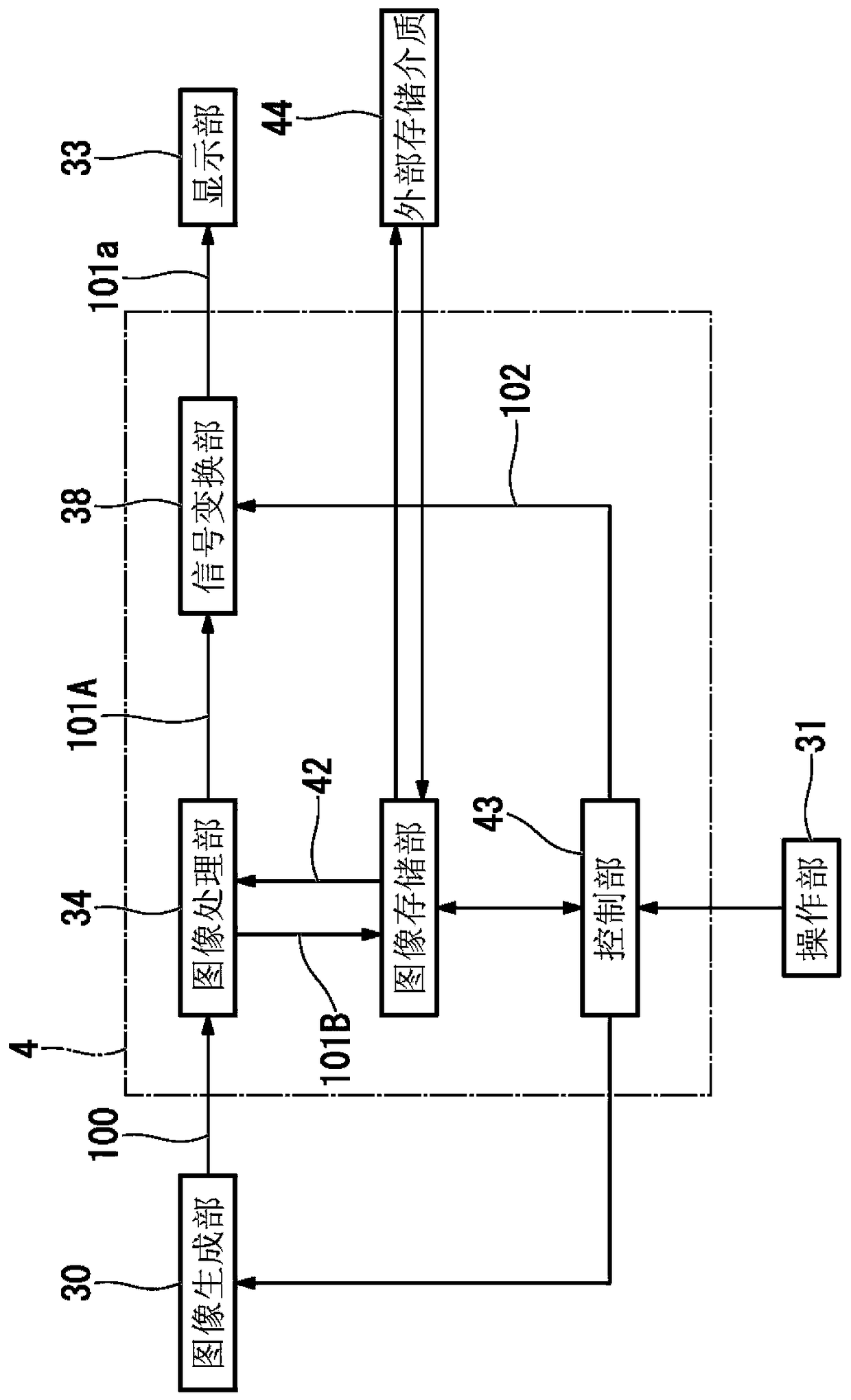
[0045] First, a first embodiment of the present invention will be described. figure 1 The appearance of the endoscope apparatus of this embodiment is shown. figure 2 The functional structure of the endoscope apparatus of this embodiment is shown. image 3 The functional structure of the control unit included in the endoscope apparatus according to this embodiment is shown.
[0046] The endoscope apparatus 1 according to this embodiment is used to take an image of a subject to be inspected, that is, an object to be inspected, and to perform measurement using the image. , or appropriately add measurement programs to enable various observations and measurements. Next, as an example of measurement, a case where stereo measurement is performed will be described.
[0047] Such as figure 1 and figure 2 As shown, an endoscope device 1 includes an optical adapter 2 for stereo measurement, an endoscope insertion unit 3, an endoscope unit 7, a CCU 9 (camera control unit), a liqu...
no. 2 approach
[0115] Next, a second embodiment of the present invention will be described. The endoscope device of the first embodiment captures images including two subject images simultaneously formed by two optical systems, but the endoscope device of the second embodiment captures images each including time-series imaging by two optical systems image of the two subjects.
[0116] In this embodiment, an optical adapter capable of switching the optical path of light incident on the imaging element is attached to the tip of the endoscope insertion portion 3 instead of the stereo measurement optical adapter 2 in the first embodiment. Figure 9 The structure of the optical adapter in this embodiment is shown. An optical adapter 21 is attached to the distal end of the endoscope insertion portion 3 . The optical adapter 21 has concave lenses 23 a , 23 b , convex lenses 24 a , 24 b , a switching unit 25 , and an imaging optical system 26 . An imaging element 3 a is arranged in the front end ...
no. 3 approach
[0146] Next, a third embodiment of the present invention will be described. In this embodiment, a case where a recorded image is reproduced to perform measurement will be described. Next, a method of making the user confirm the image used for the measurement will be described by taking the case where the measurement is performed on the personal computer 17 as an example.
[0147] For example, a file including all image information used for measurement is recorded in the personal computer 17 by the processing of ST109 (image information recording processing). At this time, information representing image information (hereinafter referred to as representative image information) that is representative of all recorded image information is also recorded in the file. In the process of ST109, the file may be recorded on an external storage medium such as a card medium connected to the endoscope apparatus 1, and the external storage medium may be connected to the personal computer 17,...
PUM
 Abstract
Description
Claims
Application Information
 - R&D
- Intellectual Property
- Life Sciences
- Materials
- Tech Scout
- Unparalleled Data Quality
- Higher Quality Content
- 60% Fewer Hallucinations
Browse by: Latest US Patents, China's latest patents, Technical Efficacy Thesaurus, Application Domain, Technology Topic, Popular Technical Reports.
© 2025 PatSnap. All rights reserved.Legal|Privacy policy|Modern Slavery Act Transparency Statement|Sitemap|About US| Contact US: help@patsnap.com


