
Anti-atherosclerosis drug efficacy evaluation method based on inflammatory responses

A technology for atherosclerosis and drug efficacy evaluation, which is applied in the fields of compound screening, material inspection products, and detection of programmed cell death.

Active Publication Date: 2014-02-05
INST OF CHINESE MATERIA MEDICA CHINA ACAD OF CHINESE MEDICAL SCI
View PDF1 Cites 3 Cited by
  • Summary
  • Abstract
  • Description
  • Claims
  • Application Information

AI Technical Summary

Problems solved by technology

[0006] Inflammation plays an important role in the whole process from the onset of AS to clinical cardiovascular events. However, the specific roles and mechanisms of many links in this complex network are still poorly understood.
In addition, although in some AS animal models, anti-inflammatory treatment targeting multiple links of inflammation has achieved certain curative effects, there is still a lack of systematic and large-scale experimental research in clinical practice.

Method used


Image

  • Anti-atherosclerosis drug efficacy evaluation method based on inflammatory responses
  • Anti-atherosclerosis drug efficacy evaluation method based on inflammatory responses
  • Anti-atherosclerosis drug efficacy evaluation method based on inflammatory responses

Examples


Embodiment 1

[0089] 1. Method for establishing EC-MC-SMC co-culture system:

[0090] 1.1 First plant SMC (HASMC) at 1×10⁵ / cm2 (volume 50 μl) on the outside of the PE membrane of Millicellinsert, turn over after 8 hours for the cells to adhere to the wall, and plant EC (HUAEC) at 1×10⁵ / cm2 on the inner side of the membrane, After co-culture for 6 days, mononuclear cells THP-1 were inoculated at 1×10⁵ / cm2, and oxLDL100μg / ml, IL-1β10ng / ml and test drug were added to culture for 24h. Pharmacodynamic experiments were carried out in this system.

[0091] 1. After 224 hours, use the tip of a pipette to gently absorb the medium in the Millicellinsert, repeat 3 times, and then absorb the supernatant (about 400 μl) into a 1ml EP tube and centrifuge at 2500rpm at 4°C for 10min. Take the supernatant and freeze it. Used to detect EC layer MCP-1, TNFα.

[0092] 1.3 The pellet is the pellet of mononuclear THP-1 cells, which is resuspended in 1ml PBS, and CD36 on the surface of monocytes is detected by...

Embodiment 2

[0104] 1 unknown drug experiment

[0105] Tanshinone IIA (Tan IIA)

[0106] The EC-SMC-MC co-culture group was used as a control, and atorvastatin was used as a positive control to detect according to the method of Example 1, and the detection results were as follows:

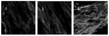
[0107] 1.1 The effect of tanshinone IIA on the secretion of MCP-1 and TNFα from EC cells The results showed that EC-SMC-MC co-cultured and stimulated with oxLDL and IL-1, ICAM-1 on the surface of EC, and TNFα, MCP- 1 was significantly increased. Tanshinone IIA can reduce the expression of ICAM-1 on the surface of EC, and the content of TNFα and MCP-1 in the co-culture supernatant, and there are significant differences compared with the model group (P<0.05 or P<0.01).

[0108] Table 5 Effect of Tanshinone IIA on the secretion of MCP-1 and TNFα from EC cells

[0109]

[0110] 1.2 The effect of tanshinone IIA on the expression of CD36 on the surface of monocytes The results showed that the expression of CD36...


PUM


Abstract

The invention discloses an anti-atherosclerosis drug efficacy evaluation method based on inflammatory responses, wherein the method comprises the following steps: step 1, planting smooth muscle cells on the outer side of a Millicell@insert PE (Poly Ethylene) membrane, and turning after the cells are adhered to the wall for 8 hours; step 2, planting endothelial cells on the inner side of the membrane, and culturing for 6 days totally; step 3, inoculating mononuclear cells THP-1, adding oxLDL, IL-1 and texted drugs, and culturing for 24 hours; step 4, detecting any one of the items of detecting contents of MCP-1 and TNFa in the endothelial cells; detecting the content of CD36 on the surface of the mononuclear cells; detecting the contents of MDA and MMP2 in the smooth muscle cells, observing the change of a smooth muscle cytoskeleton; observing the expression of ICAM-1 of the endothelial cells.

Description

Technical field: [0001] The invention relates to a drug efficacy evaluation method of a drug, in particular to an anti-atherosclerosis drug efficacy evaluation method based on an inflammatory response. Background technique: [0002] There have been several theories about the pathogenesis of atherosclerosis (AS): lipid infiltration theory, platelet aggregation and thrombosis theory, monoclonal smooth muscle cell proliferation theory, immune theory and injury response theory. The inflammation theory developed from this has explained the formation and development process of AS more comprehensively. At present, it is believed that the whole process from occurrence, development to regression of AS is a chronic inflammatory process, in which many inflammatory cells and inflammatory mediators are involved, but the mechanism of inflammation participating in the process of AS has not been fully elucidated so far. Current research mainly focuses on various inflammatory diseases. The ...

Claims


Application Information

Patent Timeline
IPC IPC(8): G01N33/68
CPCG01N33/5023G01N33/94G01N2500/10
Inventor 朱晓新李玉洁杨庆翁小刚陈颖王娅杰
Owner INST OF CHINESE MATERIA MEDICA CHINA ACAD OF CHINESE MEDICAL SCI
Learn More