Left ventricle nuclear magnetic resonance image segmentation and three-dimensional reconstruction method

A nuclear magnetic resonance image and three-dimensional reconstruction technology, applied in the field of image processing, can solve the problems of increasing the complexity of the model, the calculation amount of numerical discrete solution, and the inability to handle images with non-uniform grayscale well.

Inactive Publication Date: 2014-06-25
NORTHWESTERN POLYTECHNICAL UNIV
View PDF4 Cites 18 Cited by
  • Summary
  • Abstract
  • Description
  • Claims
  • Application Information

AI Technical Summary

Problems solved by technology

However, Chan's active contour method cannot handle images with non-uniform grayscale well, and Li's non-reinitialization method corresponds to more gradient t

Method used


Image

  • Left ventricle nuclear magnetic resonance image segmentation and three-dimensional reconstruction method
  • Left ventricle nuclear magnetic resonance image segmentation and three-dimensional reconstruction method
  • Left ventricle nuclear magnetic resonance image segmentation and three-dimensional reconstruction method

Examples


Embodiment Construction

[0050] The present invention will be further described below in conjunction with the accompanying drawings and embodiments.

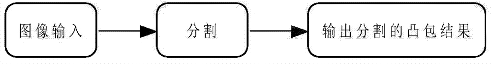
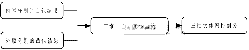
[0051] figure 1 For the flow chart of initializing and splitting modules, figure 2 It is a flowchart of the 3D reconstruction module, and the specific steps are as follows:

[0052] 1. "Initialization" step:

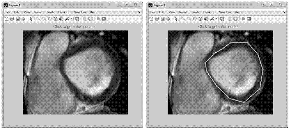
[0053] "Image input": Use RadiAnt DICOM Viewer software to convert the CMR image in dicom format into a png format image, and then read the image. image 3 The picture on the left is the png image after reading.

[0054] 2. "Split" step:

[0055] ① First establish a new variational level set model

[0056] I establish quasi-local binary simulation items for processing non-uniform gray images

[0057] Consider the grayscale value I 0 Any initial open region D in the image of , whose boundary is and D in and D out are the inner and outer regions of D. In order to implicitly track the target boundary, a level set function φ(x) can be intro...


PUM


Abstract

The invention provides a left ventricle nuclear magnetic resonance image segmentation and three-dimensional reconstruction method. According to the method, a variational level set evolution model is established to be used for segmenting the boundary of the inner membrane and the outer membrane of the left ventricle, and meanwhile, a three-dimensional curved surface and entity reconstruction scheme of the left ventricle and a corresponding mesh generation scheme are also provided. Due to the fact that a novel quasi local binary simulation item based on an edge detection operator is adopted, the method can be used for effectively segmenting images which are uneven in gray level; only the area item is considered, therefore, theoretical models involved in the method can be simplified, and the algorithm operation time of the method can also be shortened; the application of the convex hull algorithm enables the method to be capable of making up the influences such as leakage caused by fuzziness of the boundary of the outer membrane. According to the method, the three-dimensional reconstruction scheme based on the contour surface generation algorithm and secondary development and application technologies of relevant commercial software is further provided, and can help a user realize three-dimensional reconstruction of the left ventricle.

Description

technical field [0001] The invention relates to the field of image processing, in particular to a method for segmenting and reconstructing nuclear magnetic resonance images. Background technique [0002] Cardiac Magnetic Resonance (CMR) imaging can provide high-resolution images. It is one of the research hotspots in the field of medical image analysis and an important auxiliary means for the diagnosis of heart diseases. In order to make full use of the information in CMR images and provide quantitative and intuitive references for clinical diagnosis, the accurate segmentation of the intima and adventitia of the left ventricular membrane is the primary task. The left ventricular image segmentation problem mainly faces the following challenges: (1) the non-uniformity of image grayscale; (2) the adventitia border is not clear; (3) the shape of the endocardium and adventitia border in different slice images is changeable, etc. Although there are many techniques for segmenting ...

Claims


Application Information

Patent Timeline
IPC IPC(8): G06T7/00G06T17/00A61B5/055
Inventor 蔡力高昊谢文贤
Owner NORTHWESTERN POLYTECHNICAL UNIV
Why Eureka
Try Eureka