
Reduced x-ray exposure by simulating images

一种X射线、图像的技术,应用在组织消融系统领域,能够解决心律不同步、心律失常、扰乱心动周期等问题

Inactive Publication Date: 2014-07-02
BIOSENSE WEBSTER (ISRAEL) LTD
View PDF17 Cites 2 Cited by
  • Summary
  • Abstract
  • Description
  • Claims
  • Application Information

AI Technical Summary

Problems solved by technology

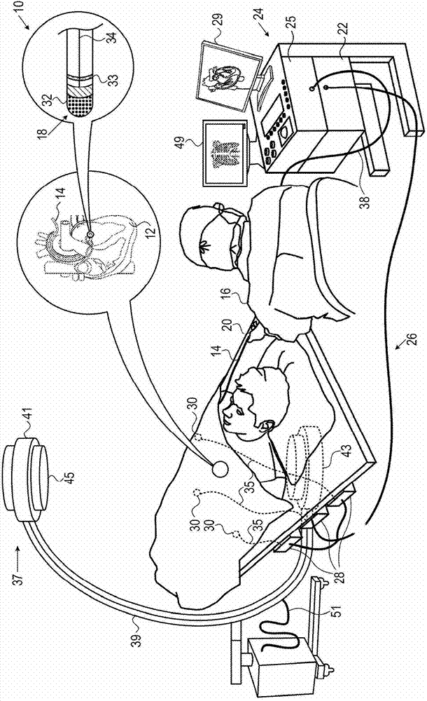
[0002] Heart rhythm disorders, such as atrial fibrillation, occur when areas of heart tissue abnormally conduct electrical signals to adjacent tissues, disrupting the normal heart cycle and causing the heart rhythms to become out of sync

Method used


Image

  • Reduced x-ray exposure by simulating images
  • Reduced x-ray exposure by simulating images
  • Reduced x-ray exposure by simulating images

Examples


example

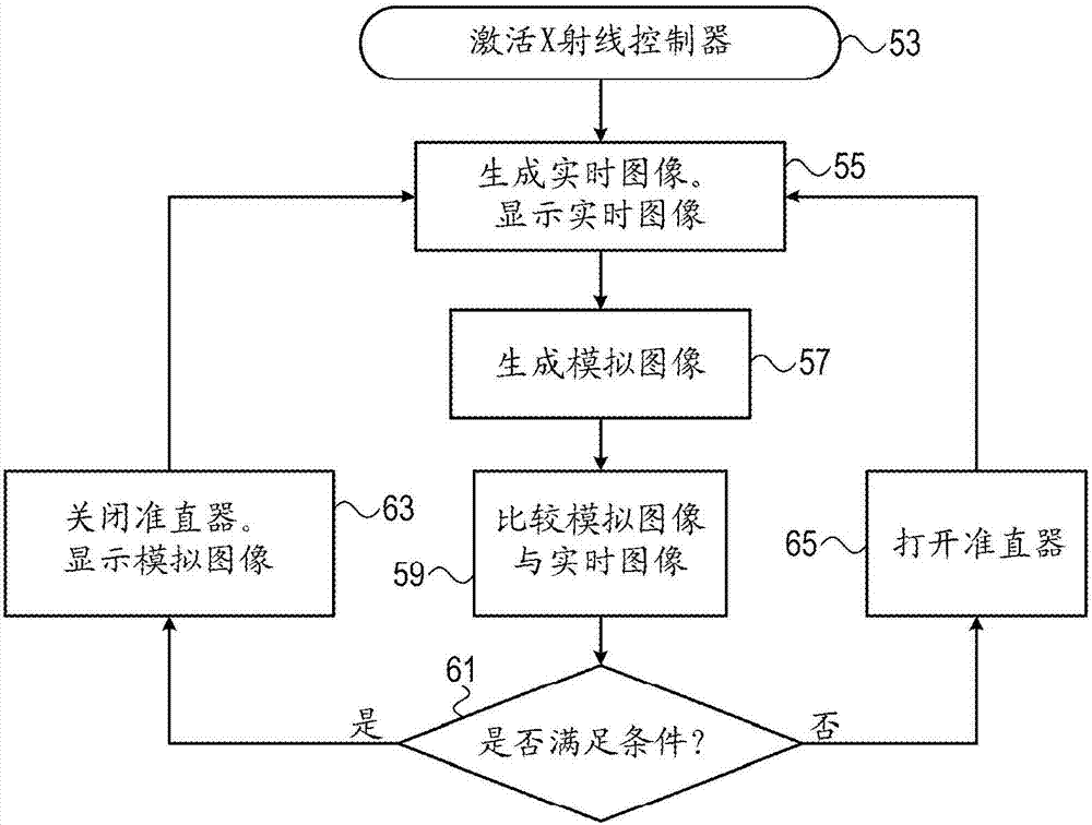
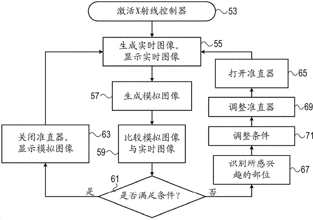
[0059] see now Figure 4 , which is based on image 3 A representative screen display of a composite or simulated image of a heart of the method described, where as a result of decision step 61 more fluoroscopic images need to be obtained. Figure 4 An image of is displayed after completing step 67. Fluoroscopy images and Carto images are presented together. The noose 75 and shaft 81 of the cardiac catheter are shown positioned in the heart as a dual image, showing their current position of the noose 75 as well as their position in previous determinations. The two rectangular regions of interest 77, 79 encompass the noose 75 and the shaft 81, respectively, but not the rest of the fluoroscopic image. Subsequent X-ray exposures will be limited to the site of interest77,79. Since the field of view of the fluoroscopic image can be limited to a specific rectangle, two sites of interest 77, 79 can be acquired at two different points in time.

[0060] It has been found that usin...


PUM


Abstract

Medical imaging is carried out by transmitting X-ray emissions through a subject, generating a first set of temporally separated images of an area of interest in the subject from the transmitted X-ray emissions, generating a second set of temporally separated images from the area of interest from a second source, and combining the sets to produce combined images of the area of interest. The method is further carried out by blocking the X-ray emissions from reaching the subject, updating the second set, combining the first set with the updated second set to produce updated combined images, displaying the updated combined images, and when a predetermined condition is satisfied, iterating updating the second set, combining the first set with the updated second set, and displaying the updated combined images.

Description

technical field [0001] The present invention relates to tissue ablation systems. More specifically, the present invention relates to tissue ablation systems that include a combination of fluoroscopic and non-fluoroscopic imaging techniques. Background technique [0002] Arrhythmias such as atrial fibrillation occur when parts of the heart tissue conduct electrical signals abnormally to adjacent tissues, disrupting the normal heart cycle and causing the heart rhythms to become out of sync. [0003] Surgery to treat cardiac arrhythmias involves surgically disrupting the source of the signal causing the arrhythmia, as well as disrupting the conduction pathways for such signals. Ablation methods disrupt unwanted electrical pathways by creating nonconducting lesions. Contents of the invention [0004] More recently, sophisticated electroanatomical mapping systems have been used to detect arrhythmogenic regions within the heart and to guide ablation. Fluoroscopy or computed t...

Claims


Application Information

Patent Timeline
Patent Type & Authority Applications(China)
IPC IPC(8): A61B6/00A61B18/12
CPCA61B6/54A61B6/503A61B18/1492A61B6/463A61B2018/00577A61B5/0402A61B6/06A61B6/487A61B5/062A61B6/5247A61B5/6876A61B6/4441A61B2019/5238A61B6/542A61B6/5288A61B5/0538A61B5/063A61B5/0035A61B19/5244A61B6/52A61B6/4225A61B6/12A61B5/042A61B2017/00026A61B2017/00088A61B2017/00092A61B2018/00357A61B2090/376A61B2034/2051A61B5/318
Inventor M.阿米特
Owner BIOSENSE WEBSTER (ISRAEL) LTD
Features
  • Generate Ideas
  • Intellectual Property
  • Life Sciences
  • Materials
  • Tech Scout
  • Unparalleled Data Quality
  • Higher Quality Content
  • 60% Fewer Hallucinations
Learn More