Multi-energy-spectrum CT imaging method and imaging system

A CT imaging and multi-energy spectrum technology, applied in the field of radiation imaging, can solve the problem of low reconstruction quality

Active Publication Date: 2014-07-02
CAPITAL NORMAL UNIVERSITY +1
View PDF7 Cites 32 Cited by
  • Summary
  • Abstract
  • Description
  • Claims
  • Application Information

AI Technical Summary

Problems solved by technology

[0004] The purpose of the present invention is to solve the problem of low reconstruction quality existing in the existing multi-energy spectrum CT image reconstruction process, and to provide a multi-energy spectrum CT imaging method and imaging system that can accurately realize image reconstruction

Method used


Image

  • Multi-energy-spectrum CT imaging method and imaging system
  • Multi-energy-spectrum CT imaging method and imaging system
  • Multi-energy-spectrum CT imaging method and imaging system

Examples


Embodiment Construction

[0076] The above and other technical features and advantages of the present invention will be further described below in conjunction with the accompanying drawings.

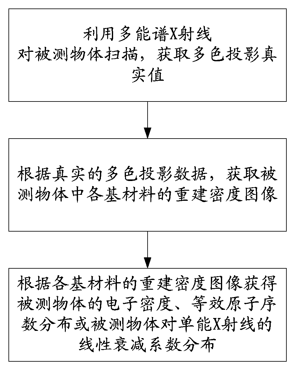
[0077] see figure 1 , is a flow chart of the multi-energy spectral CT imaging method proposed by the present invention. Depend on figure 1 It can be seen that the multi-energy spectral imaging method of the present invention comprises the following steps:

[0078] S1: Use multi-energy spectrum X-rays to scan the measured object to obtain the true value of multi-color projection;

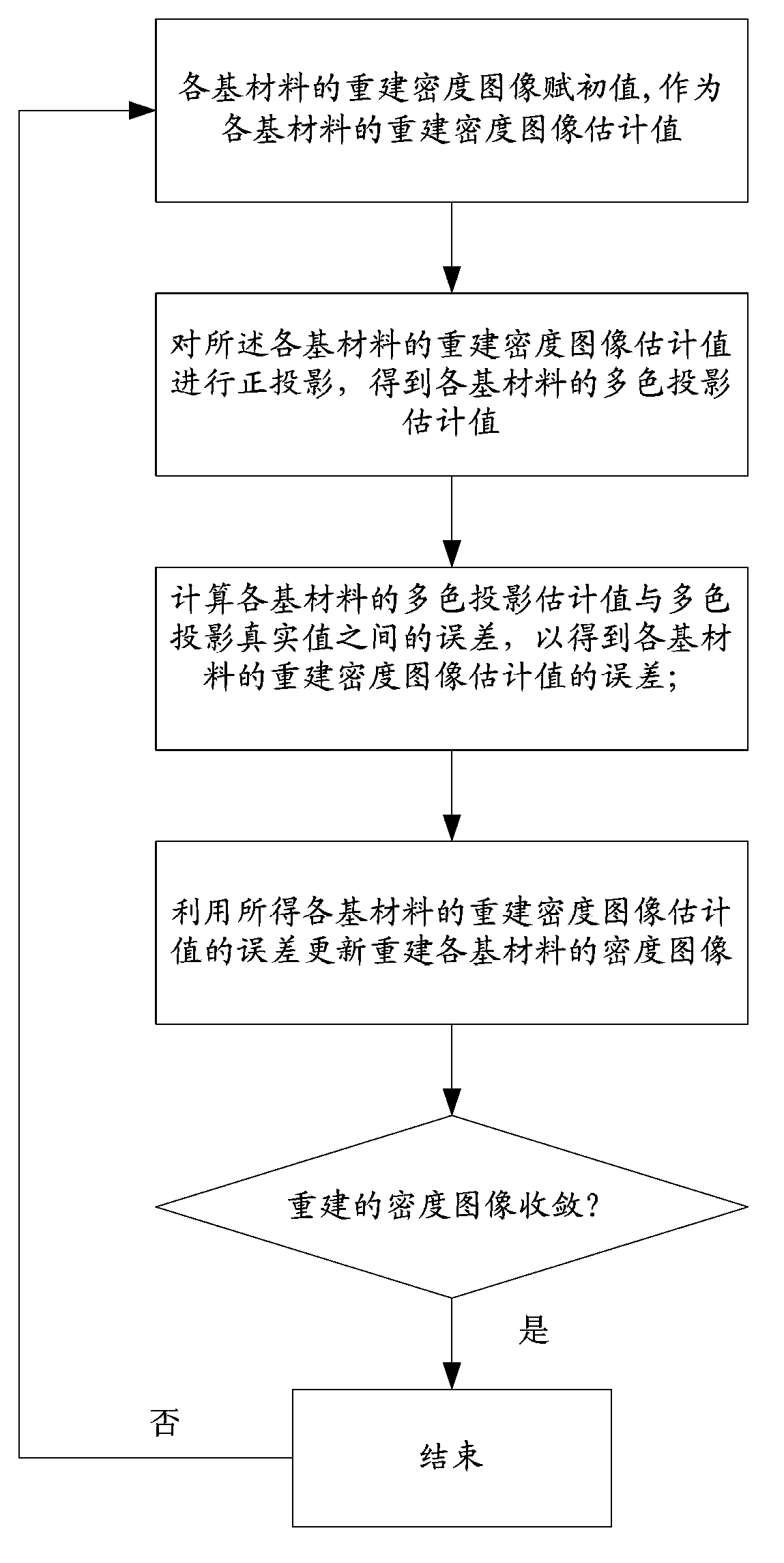
[0079] S2: Obtain the reconstructed density image of each base material in the object under test according to the real multi-color projection data;

[0080] S3: Obtain the electron density and equivalent atomic number distribution of the measured object or the linear attenuation coefficient distribution of the measured object for single-energy X-rays according to the reconstructed density images of each base material.

[0081] The m...


PUM


Abstract

The invention discloses a multi-energy-spectrum CT imaging method and an imaging system. The multi-energy-spectrum CT imaging method comprises the following steps: scanning the object to be detected, and reconstructing the density image of a basic material so as to finally obtain the electron density and equivalent atomic number distribution of the object to be detected or linear attenuation coefficient distribution of the object under the radiation of mono-energetic X-rays. The multi-energy-spectrum CT reconstruction method comprises the following steps: assigning an initial value for the density image of a basic material to be reconstructed; carrying out orthographic projection on an estimated image so as to obtain a projection estimated value; estimating the error of the reconstructed image; updating the image by utilizing the estimated error of the reconstructed image, and repeating the steps mentioned above until the reconstructed density image is convergent. The multi-energy-spectrum CT imaging system comprises a scanning module, a reconstruction module, and a statistics module, wherein the reconstruction module comprises an initializing module, a projecting module, a correcting module, and an updating module.

Description

technical field [0001] The invention relates to the field of radiation imaging, in particular to a multi-energy spectral X-ray CT imaging method. Background technique [0002] Traditional X-ray CT uses an X-ray energy spectrum to scan the object under test. The CT value of the reconstructed image is related to the material and density of the measured object, as well as the energy spectrum of X-rays. Therefore, reconstructed images of different substances may have the same or similar CT values. This makes it impossible for traditional CT to distinguish them. However, multispectral CT uses multiple energy spectra to scan the object under test, so more information about the object under test can be obtained. The information can be used to reconstruct the equivalent atomic number and electron density image of the measured object, or to reconstruct the density image of the base material. Multispectral CT has broad application prospects in bone density measurement, PET attenua...

Claims


Application Information

Patent Timeline
Patent Type & Authority Applications(China)
IPC IPC(8): G01N9/24A61B6/03
Inventor 张朋赵云松张慧滔
Owner CAPITAL NORMAL UNIVERSITY
Why Eureka
Try Eureka