Multimodal multi-dimensional blood vessel fusion method and system

A fusion method and multi-dimensional technology, applied in the field of medical device data image processing, can solve the problems of insecure stability and registration accuracy, failure to discard other information of blood vessels, failure to provide blood vessel display, etc., to improve stability and registration The effect of precision

Inactive Publication Date: 2014-07-23
CREALIFE MEDICAL TECH BEIJING
View PDF4 Cites 26 Cited by
  • Summary
  • Abstract
  • Description
  • Claims
  • Application Information

AI Technical Summary

Problems solved by technology

[0003] However, the traditional blood vessel fusion method is based on image fusion, that is, two images are fused first and then blood vessels are extracted. This method cannot discard other information

Method used


Image

  • Multimodal multi-dimensional blood vessel fusion method and system
  • Multimodal multi-dimensional blood vessel fusion method and system
  • Multimodal multi-dimensional blood vessel fusion method and system

Examples


Embodiment Construction

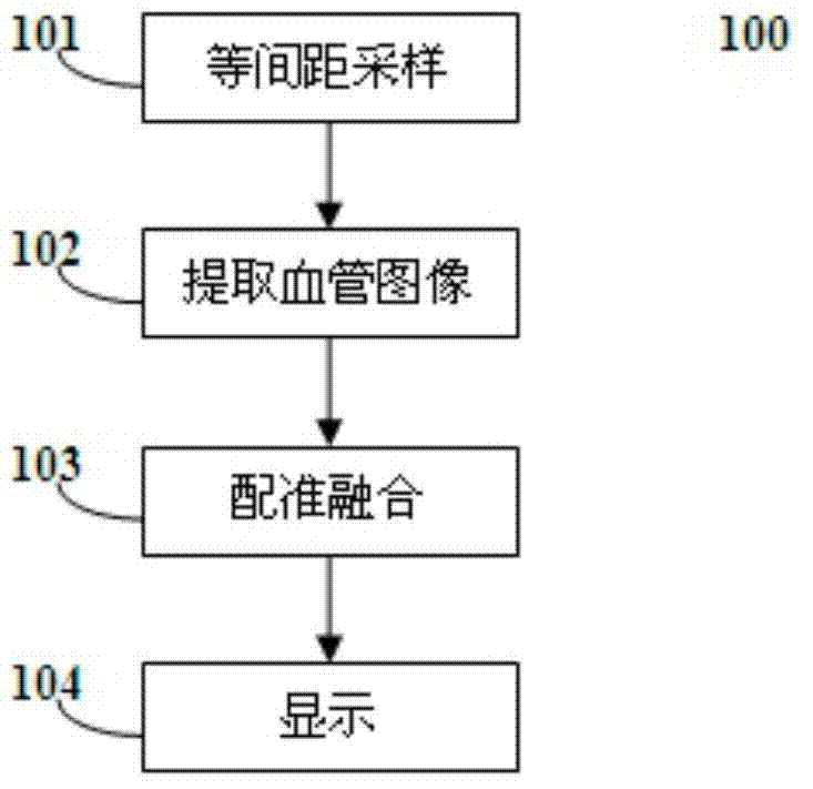
[0023] The overall process of the present invention is as figure 1 As shown, the specific steps are as follows:

[0024] Execute step 101 for equidistant sampling, perform equidistant sampling on multiple original images of the medical images, obtain multiple medical images with the same resolution, perform step 102 to extract blood vessel images, and extract blood vessel images in the multiple medical images respectively, And extract the blood vessel center line corresponding to the blood vessel image, perform step 103 registration and fusion, and carry out registration and fusion on the blood vessel center line through the ICP (Iterative Closest Point, iterative closest point algorithm) point set registration algorithm to obtain three-dimensional data, Step 104 is executed to display the three-dimensional data synchronously in the form of cross-sectional, coronal and sagittal tomographic images. Wherein step 102 also includes eliminating other images in the medical image ex...


PUM


Abstract

The invention discloses a multimodal multi-dimensional blood vessel fusion method and a multimodal multi-dimensional blood vessel fusion system, and relates to medical equipment data image processing. The method comprises the steps of respectively extracting blood vessel images in a plurality of medical images, and extracting blood vessel center lines corresponding to the blood vessel images; registering and fusing the blood vessel center lines through an iterated near point algorithm, so as to obtain three-dimensional data; and synchronously displaying the three-dimensional data in the way of the cross-sectional image of cross sections, coronal planes and vertical planes. According to the method, other information outside the blood vessels can be rejected, so that no other covers can be generated when a doctor observes the blood vessels.

Description

technical field [0001] The invention relates to medical device data image processing, in particular to a multi-modal and multi-dimensional blood vessel fusion method and system. Background technique [0002] In the process of diagnosing vascular diseases through medical images, medical images of different modalities and dimensions can reflect different vascular structures and lesion information. For example, DSA (Digital Subtraction Angiography) can clearly display the vascular structure at all levels, and can also record the hemodynamic characteristics of blood vessels in real time; CTA (CT Angiography) can obtain 3D stereoscopic images through 3D reconstruction through computer screens. The upper multi-angle rotation can provide a comprehensive three-dimensional shape of the malformed blood vessel, the details of the anatomical structure of the perivascular tissue, and guide the design of the surgical approach; the imaging resolution and definition of MRA (magnetic resonan...

Claims


Application Information

Patent Timeline
IPC IPC(8): G06T5/50
Inventor 李晓光王民汉周宇
Owner CREALIFE MEDICAL TECH BEIJING
Why Eureka
Try Eureka