Whole heart extracting method based on heart CT image

A CT image and extraction method technology, applied in image enhancement, image analysis, image data processing and other directions, can solve the problems of residual noise tissue, loss of cardiac edge tissue, etc., to achieve accurate extraction effect, strong adaptability, and high operating efficiency Effect

Inactive Publication Date: 2014-08-13
SHENZHEN GRADUATE SCHOOL TSINGHUA UNIV
View PDF2 Cites 10 Cited by
  • Summary
  • Abstract
  • Description
  • Claims
  • Application Information

AI Technical Summary

Problems solved by technology

[0004] The above two types of methods start from the problem itself, directly take the target area of ​​the heart as the research object, and expect to use various methods to extract the entire heart from the entire CT tomographic image, but often cause problems such as loss of heart edge tissue or residual noise tissue

Method used


Image

  • Whole heart extracting method based on heart CT image
  • Whole heart extracting method based on heart CT image
  • Whole heart extracting method based on heart CT image

Examples


Embodiment Construction

[0026] The present invention will be further described below in conjunction with the accompanying drawings and embodiments. These more detailed descriptions are intended to help the understanding of the present invention, but should not be used to limit the present invention. From the present disclosure it will be apparent to those skilled in the art that the present invention may be practiced without some or all of these specific details. In other instances, well known operating procedures have not been described in detail in order not to obscure the invention.

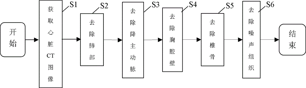
[0027] This protocol starts from a reverse perspective, takes the non-target area as the research object, and achieves the purpose of extracting the whole heart by gradually removing non-cardiac tissues such as the chest wall, lungs, vertebrae, and descending aorta.

[0028] Such as figure 1 As shown, this whole heart extraction method based on cardiac CT image comprises the following steps:

[0029] Step S1, acqu...


PUM


Abstract

A whole heart extracting method based on a heart CT image comprises the steps of obtaining the heart CT image, removing the lung part, the descending aorta part, the thoracic cavity wall part and the vertebra part of the heart CT image to obtain a middle image, and removing the noise part in the middle image. The whole heart extracting method based on the heart CT image gradually removes the non-heart parts such as the thoracic cavity wall, the lung, the vertebra and the descending aorta in an reverse angle mode to achieve the purpose of extracting the whole heart, has the advantages of being high in self-adaptation performance, high in operation efficiency, accurate in extraction effect and the like, can be rapidly embedded in an existing medical network, and achieves remote auxiliary diagnosis.

Description

technical field [0001] The invention belongs to the field of computer vision and image processing, in particular to a whole heart extraction method based on heart CT images. Background technique [0002] Whole heart extraction has important application value in many fields, for example, in the diagnosis of cardiovascular diseases, it provides three-dimensional visualization of coronary arteries; when making radiotherapy plans for lung tumors, it assists radiotherapists in delineating radiotherapy target areas, avoiding healthy tissues such as the heart ; and separate diagnosis of the ventricle, atrium and other chambers of the whole heart, etc. [0003] At present, most studies on heart extraction from CT images use the method of directly extracting the target area of ​​the heart, which is mainly realized by using the high contrast between the blood pool in the heart chamber and other tissues after injection of contrast agent. The methods are roughly divided into two catego...

Claims


Application Information

Patent Timeline
Patent Type & Authority Applications(China)
IPC IPC(8): G06T7/00G06T5/00
Inventor 袁克虹张硕张广
Owner SHENZHEN GRADUATE SCHOOL TSINGHUA UNIV
Why Eureka
Try Eureka