Method for visible serial segmentation of human body slice images based on skeleton angular points

A technology of image sequence and slicing, applied in image analysis, image data processing, instruments, etc., to achieve good robustness, low time complexity, and less manual intervention

Active Publication Date: 2014-08-27
DALIAN UNIV OF TECH
View PDF2 Cites 13 Cited by
  • Summary
  • Abstract
  • Description
  • Claims
  • Application Information

AI Technical Summary

Problems solved by technology

[0007] The purpose of the present invention is to meet the needs of visible human body research, solve the problem of fast and accurate segmentation of main organs in visible human body slice images, and thus propose an automatic serialized color image segmentation method for human body slices

Method used


Image

  • Method for visible serial segmentation of human body slice images based on skeleton angular points
  • Method for visible serial segmentation of human body slice images based on skeleton angular points
  • Method for visible serial segmentation of human body slice images based on skeleton angular points

Examples


Embodiment Construction

[0037] In order to make the purpose, technical solution and advantages of the present invention clearer, the present invention will be further described in detail below in conjunction with the accompanying drawings and embodiments.

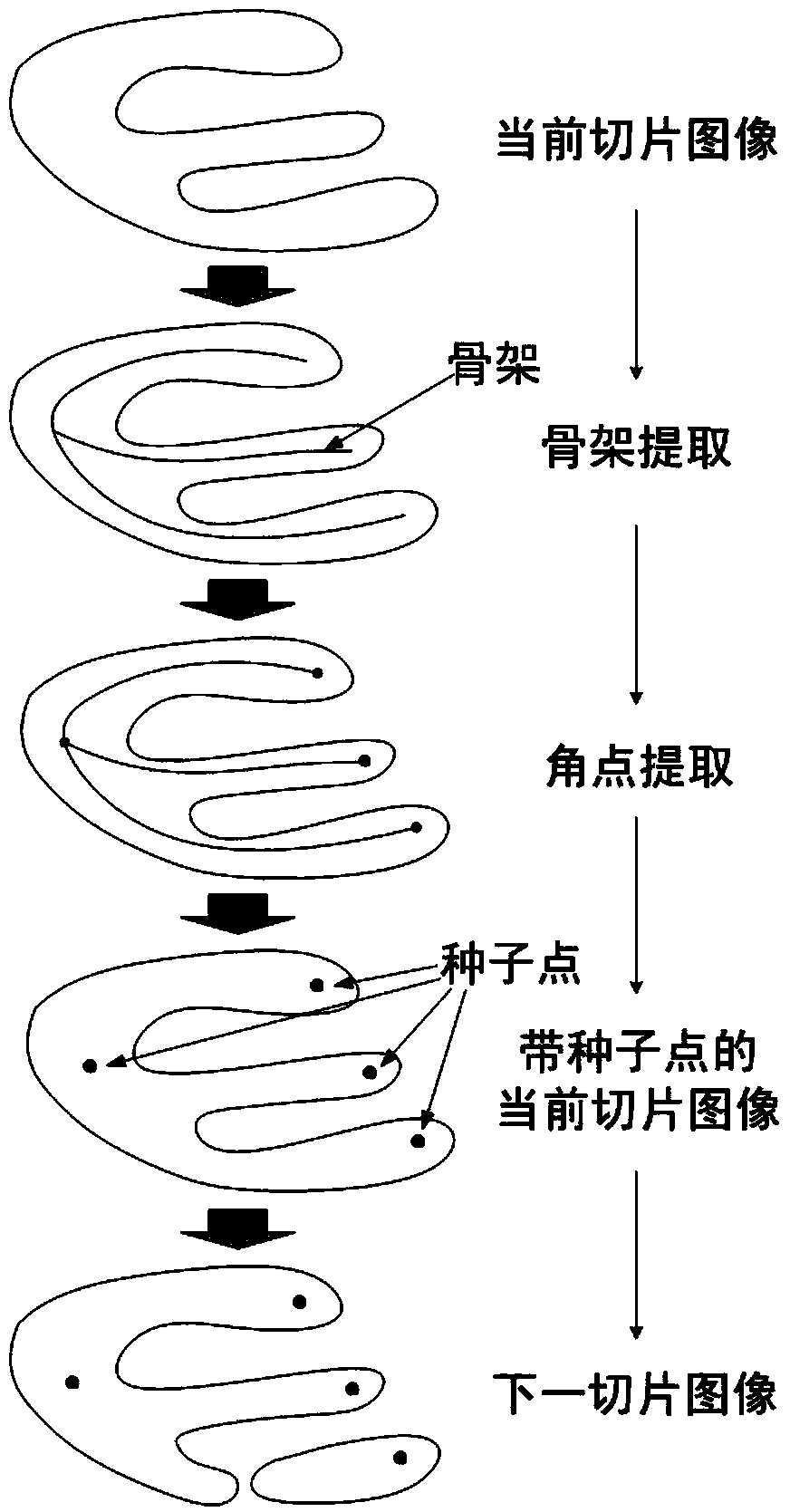
[0038] In order to quickly and accurately segment the main organs of the visualized human body slices, the present invention proposes an automatic sequential color image segmentation method, see image 3 , see the description below:

[0039] A method for automatically serializing color image segmentation, the method comprising the following steps:

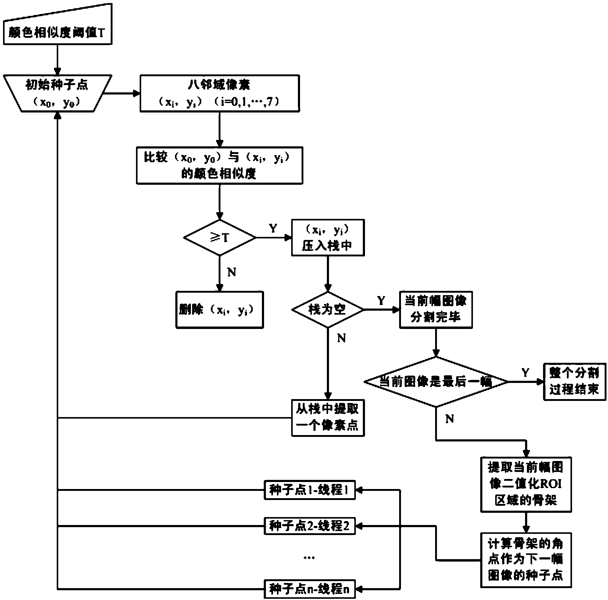
[0040] (1) Load the serialized visible human (Visible Human) color slice image dataset and the spare adjacent color pixel color similarity threshold;

[0041] (2) open up the seed point set space, and manually select some seed points on the first image, and store them in the seed point set space;

[0042](3) Comparison of color similarity between adjacent pixels: extract a seed point from the seed po...


PUM


Abstract

Disclosed is a method for visible serial segmentation of human body slice images based on skeleton angular points. The method comprises the steps of loading a serial colored slice data set and a color similarity threshold value; developing a seed point set, selecting multiple seed points manually, and storing the seed points in the seed point set; calculating the color similarity between each seed point and a neighboring pixel, storing a mark if the color similarity is larger than the similarity threshold value, discarding the mark otherwise, and taking the obtained seed point set as the target binary image of the current image; ending the process if the current image is the last one; conducting skeleton extraction on the target binary image to obtain a target skeleton image, and emptying the seed point set; acquiring multiple angular points of the skeleton image with the angular point detection method to serve as the seed point of the next image, and storing the seed points in the seed point set; traversing the image sequence to achieve continuous segmentation until all the images to be segmented are segmented. By means of the method, a target human organ can be obtained from a three-dimensional colored slice image data set continuously and automatically through segmentation.

Description

technical field [0001] The invention belongs to the technical field of human organ visualization, and in particular relates to a method for sequentially segmenting visualized human body slice images based on skeleton corner points. Background technique [0002] Human organ visualization research emerged in the late 20th century, based on information technology and medical science, it is an advanced and interdisciplinary research field. Obtaining and digitizing the basic data of human organ structure is the basis and premise of scientific research on human body visualization, which has high academic significance and research value. At present, the human body visualization research has completed the stage of accurate acquisition of ultra-large-scale (hundred GB level) color slice images. Faced with such huge image data, it is a huge project to extract the geometric model of human organs using heavy manual segmentation or semi-automatic interactive segmentation methods. There...

Claims


Application Information

Patent Timeline
Patent Type & Authority Applications(China)
IPC IPC(8): G06T7/00
Inventor 刘斌田博范珏辉王蒙
Owner DALIAN UNIV OF TECH
Why Eureka
Try Eureka