Full-automatic CT image kidney segmentation method

A CT image, fully automatic technology, applied in the field of image processing, can solve problems such as indivisibility, achieve the effect of reducing workload, improving efficiency and accuracy, and avoiding errors

Inactive Publication Date: 2014-11-19
SOUTHEAST UNIV
View PDF2 Cites 9 Cited by
  • Summary
  • Abstract
  • Description
  • Claims
  • Application Information

AI Technical Summary

Problems solved by technology

[0003] Technical problem to be solved: Aiming at the deficiencies in the prior art, the present invention proposes a fully automatic CT image kidney segmentation method, which provides a method for computer automatic segmentation of kidney CT images, a...

Method used


Image

  • Full-automatic CT image kidney segmentation method
  • Full-automatic CT image kidney segmentation method
  • Full-automatic CT image kidney segmentation method

Examples


Embodiment Construction

[0041] The present invention will be further described below in conjunction with the accompanying drawings.

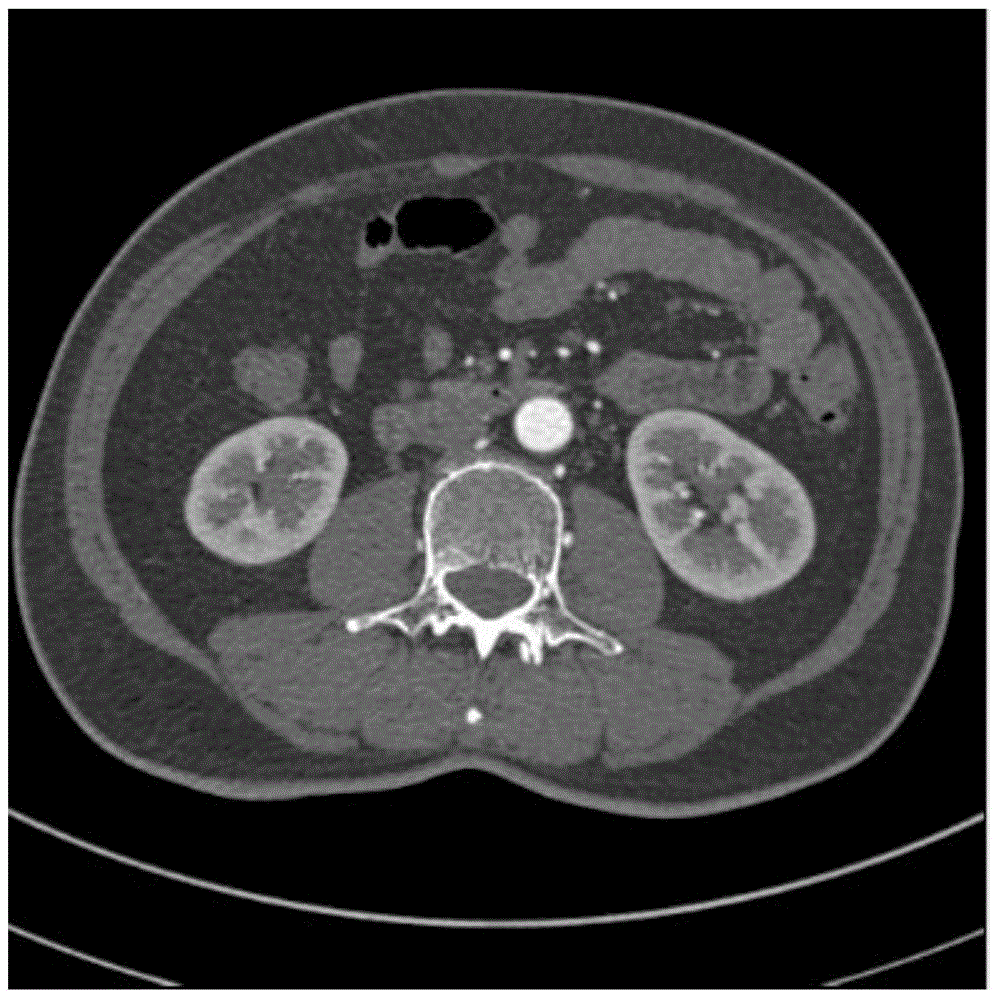
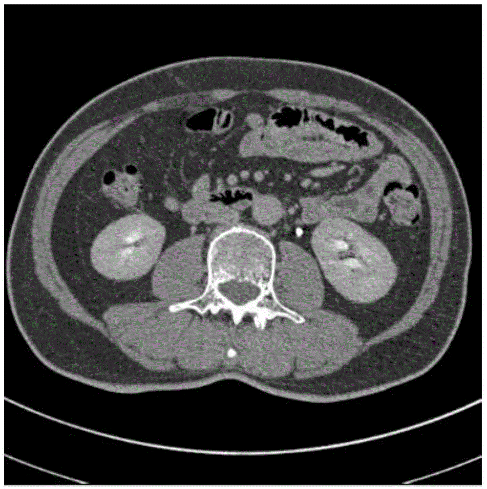
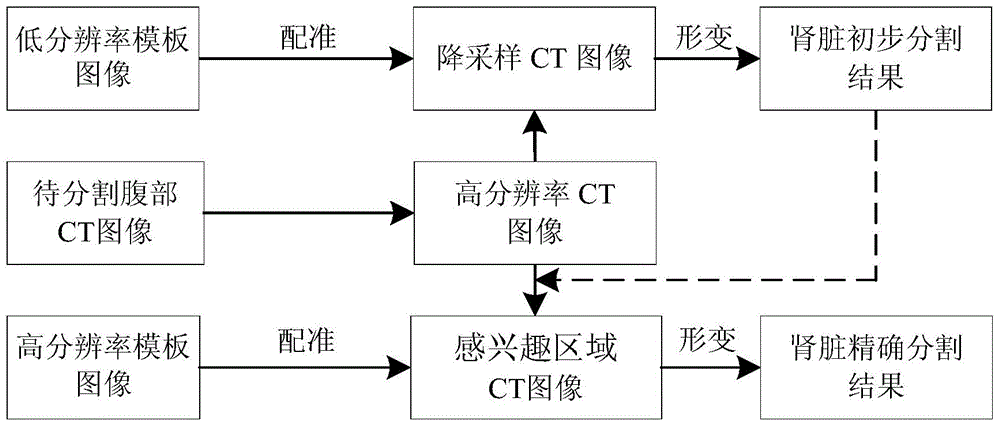
[0042] Such as image 3 Shown is the flowchart of the present invention. Including the following steps:

[0043] Step (1), the method in the present invention depend on the establishment of expert library: first select template CT image, then the template CT image that is selected is reconstructed into each direction of X, Y, Z in the three-dimensional Cartesian coordinate system by image linear interpolation algorithm Volume data with equal resolution; then the experts manually draw the kidney boundary and mark the kidney area in this volume data, and generate the expert labeling result of the kidney area; finally, the volume data and the kidney area marked by the expert are respectively Downsampling, the downsampling rate is set to 4, so that a low-resolution template image with a resolution of 1 / 64 of the image size before downsampling can be generated, and N=8 lo...


PUM


Abstract

The invention provides a full-automatic CT image kidney segmentation method. Multi-template-based image matching algorithm is used and a multi-resolution two-step algorithm strategy is adopted. A region of interest of the kidney in a low-resolution CT image is firstly positioned, and then kidney tissues in a region of interest in a high-resolution CT image are precisely segmented. The experimental result shows that in view of renal arteriography and urography CT images, accurate segmentation results can be provided, the workload of manual segmentation by a doctor can be greatly reduced, and the efficiency and effects of diagnosis and treatment can be effectively improved.

Description

technical field [0001] The invention relates to the technical field of image processing, in particular to a fully automatic CT image kidney segmentation method. Background technique [0002] CT images have been widely used in the clinical diagnosis of renal tumors, kidney stones and other urological diseases. By injecting contrast agent and adjusting image acquisition parameters during CT image acquisition, the kidney, renal tumor, renal artery, renal cortex, renal medulla, ureter and other tissues can be displayed to obtain anatomical and physiological parameters for disease diagnosis. In addition, with the widespread development of minimally invasive interventional surgery for kidney cancer, kidney stones and other diseases, this information can also provide a reliable basis for preoperative planning of minimally invasive surgery. In order to accurately obtain these parameters, it is usually necessary to segment the kidney area in the CT image. If only relying on the doct...

Claims


Application Information

Patent Timeline
IPC IPC(8): G06T7/00
Inventor 杨冠羽王征吕力兢顾金金舒华忠
Owner SOUTHEAST UNIV
Why Eureka
Try Eureka