Nuchal translucency image segmentation method, device and system

An image segmentation and transparent layer technology, which is applied in the field of medical image technology processing, can solve the problems of long time-consuming fetal ultrasound image training and learning, difficult to obtain training sets, etc., and achieve the effect of intuitive observation and short time-consuming

Active Publication Date: 2014-11-19
SONOSCAPE MEDICAL CORP
View PDF3 Cites 27 Cited by
  • Summary
  • Abstract
  • Description
  • Claims
  • Application Information

AI Technical Summary

Problems solved by technology

Chinese patent application No. 201310024351.1 proposes a Bayesian classifier-based automatic measurement of fetal cervical translucency, but this measurement

Method used


Image

  • Nuchal translucency image segmentation method, device and system
  • Nuchal translucency image segmentation method, device and system
  • Nuchal translucency image segmentation method, device and system

Examples


Embodiment 1

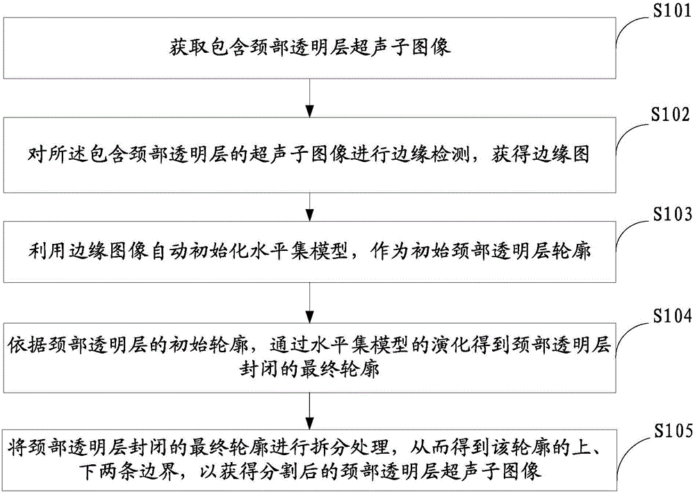
[0030] Such as figure 1 As shown, the fetal neck translucency segmentation method comprises the steps:

[0031] S101. Acquire an ultrasound sub-image including a fetal cervical translucency.

[0032] First, the ultrasonic diagnostic system processes the signal after receiving the external signal to generate an ultrasonic image including the cervical translucency; then, place the cervical translucency in the middle of the image, and determine it with a trackball, mouse or button Including the approximate range of the cervical translucency, the ultrasonic image is clipped to obtain a rough ultrasound sub-image of the target cervical translucency, which not only reduces the interference information, but also enhances the real-time performance of the algorithm.

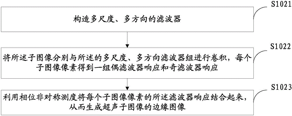
[0033] S102. Perform edge detection on the ultrasonic sub-image containing the cervical translucency to obtain an edge map.

[0034] Canny, sobel and other gradient-based methods can be used for edge detection, but thes...

Embodiment 2

[0077] Based on the segmentation of the cervical translucency, its thickness can also be measured. That is, the thickness of the neck transparent layer is found by calculating the Euclidean distance between the contour points of the upper boundary and the lower boundary.

[0078] Calculate the distance between the upper and lower boundary contour points: D i = ( x i 1 - x i 2 ) 2 + ( y i 1 - y i 2 ) 2 ...

Embodiment 3

[0082] Such as Figure 5 As shown, the present invention also provides an apparatus 300 capable of implementing the method described in Embodiment 1.

[0083] The apparatus 300 includes: an acquisition module 301 , a first calculation module 302 , a second calculation module 303 , a third calculation module 304 , and a fourth calculation module 305 .

[0084] An acquisition module 301, configured to acquire a sub-image including the cervical translucency layer.

[0085] The first calculation module 302 is configured to obtain an edge map of the sub-image through an edge detection function based on a phase asymmetric feature.

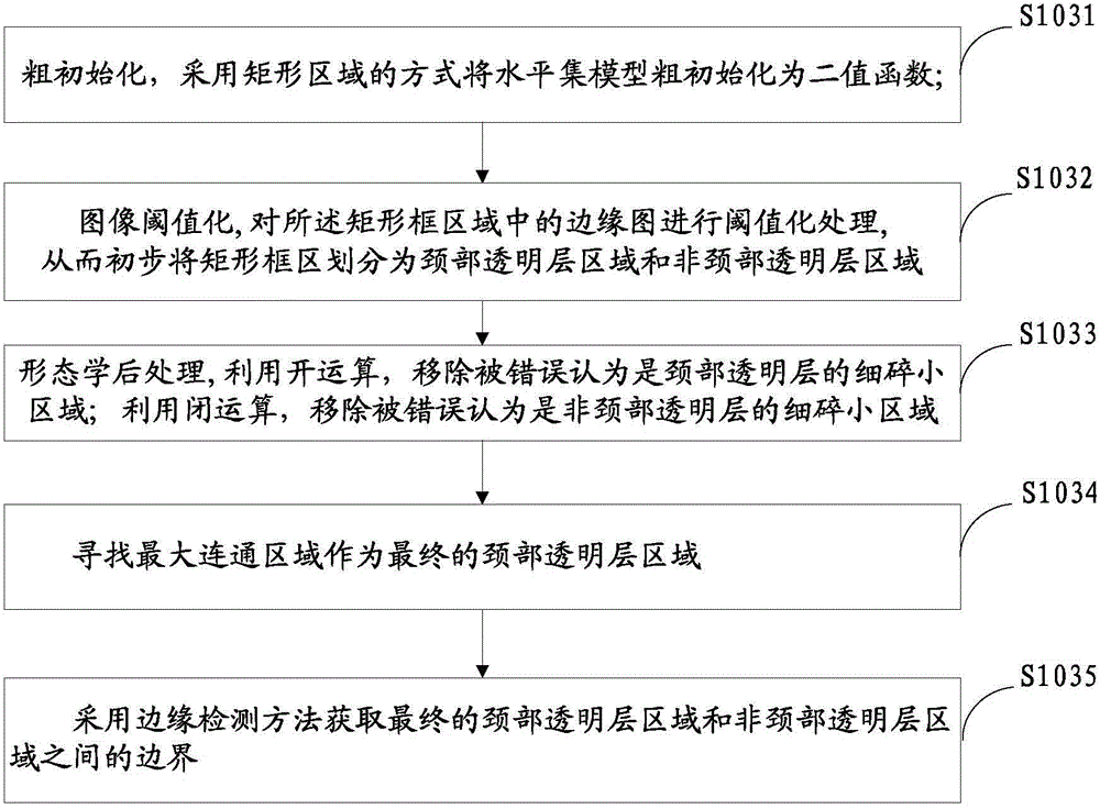
[0086] The second calculation module 303 is configured to use the edge image to automatically initialize the level set model as the initial contour of the transparent layer of the neck.

[0087] The third calculation module 304 is configured to obtain the final contour closed by the cervical translucency through the evolution of the level set model acc...


PUM


Abstract

The invention provides a nuchal translucency segmentation method. Firstly, an edge image is adopted to automatically initiate the nuchal translucency contour; then, a level set model is used for pushing evolution of the initial contour, and a closed final contour of the nuchal translucency is acquired; and finally, splitting is carried out on the closed final contour of the nuchal translucency, an upper boundary and a lower boundary of the contour are obtained, and a nuchal translucency ultrasonic sub image after segmentation is obtained. In addition, the invention also provides a device and a system related with the method. Thus, segmentation and measurement can be effectively and quickly carried out on the nuchal translucency.

Description

technical field [0001] The invention relates to the field of medical image technology processing, in particular to a method, device and system for segmenting images of a fetal neck transparent layer. Background technique [0002] Nuchal Translucency (NT) ultrasonography is the preferred method for prenatal screening of fetal abnormalities in early pregnancy, especially in the screening of fetal chromosomal abnormalities. The thickness of fetal nuchal translucency Thickening has a higher sensitivity to fetal abnormalities. The measurement of the thickness of the fetal cervical translucency in early pregnancy has very important clinical diagnostic value in detecting fetal abnormalities, which is conducive to guiding the prognosis of the fetus and improving prenatal and postnatal care. [0003] In the current measurement of the thickness of the cervical translucency in fetal ultrasound images, manual measurement is the main method. Since the manual measurement of NT thickness...

Claims


Application Information

Patent Timeline
IPC IPC(8): G06T7/00A61B8/00
Inventor 高梁冯乃章朱勇
Owner SONOSCAPE MEDICAL CORP
Why Eureka
Try Eureka