Imaging method for MRI contrast enhancement

An imaging method and contrast technology, applied in the field of magnetic resonance imaging and medical imaging, can solve problems such as adverse allergic reactions, and achieve the effect of solving the problem of excessive scanning time

Active Publication Date: 2014-11-26
SUZHOU INST OF BIOMEDICAL ENG & TECH CHINESE ACADEMY OF SCI
View PDF4 Cites 19 Cited by
  • Summary
  • Abstract
  • Description
  • Claims
  • Application Information

AI Technical Summary

Problems solved by technology

However, this imaging enhancement method has certain safety issues. For example, although gadolinium contrast agents for magnetic resonance imaging have been proven to be safer in clinical use compared with iodine contrast agents for X-ray imaging or CT imaging, but There are still 0.03-0.1% adverse allergic reactions

Method used


Image

  • Imaging method for MRI contrast enhancement
  • Imaging method for MRI contrast enhancement
  • Imaging method for MRI contrast enhancement

Examples


Embodiment Construction

[0031] The present invention will be described in detail below with reference to the accompanying drawings and in combination with embodiments.

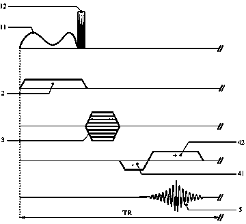
[0032] refer to figure 1 and figure 2 As shown, the specific steps are:

[0033] Step 1: Fix the two kinds of biological tissue samples in the central detection area of ​​the magnetic resonance imager, and measure the T 1 and T 2 relaxation time;

[0034] Step 2: At the same time when layer selection gradient 2 is turned on, the optimized inversion pulse 11 and the 90° excitation pulse 12 are sequentially applied to the biological tissue sample; the optimized inversion pulse 11 is a key component of the present invention, and numerical optimization based on gradient ascent can be used The method is obtained, and the specific description is as follows: Based on the T of different tissues obtained in step 1 1 and T 2 For relaxation time, the α-β model of the uncoupled two-spin system is established, which is characterized ...


PUM


Abstract

The invention discloses an imaging method for MRI contrast enhancement. Optimized inversion pulses are utilized to replace inversion pulses of a conventional inversion recovery sequence in MRI and to restore delay time, under the precise control of the optimized pluses, spin of different tissue will be evolved towards the trend of maximization of longitudinal magnetic moment differences, and the maximum longitudinal magnetic moment difference is obtained at the end moment of the pluses; on that basis, 90-degree excitation read pluses are applied to enable the maximized magnetic moment difference among the tissue to be turned over to a transverse plane, gradient echo signals are collected to form k spatial data, and the purpose of contrast enhancement among the tissue can be finally achieved through improvement on a phase sensitive image reconstruction method. According to the imaging method, the problem of too long scanning time of the conventional inversion recovery sequence is solved, the advantages of flexibility of optimized pulse waveforms and flexibility of the phase sensitive image reconstruction method are fully utilized, use of expensive magnetic resonance contrast media can be avoided, and the imaging method is superior to an existing MRI contrast enhancement method both in performance and in cost.

Description

technical field [0001] The invention relates to the field of medical imaging, in particular to the field of magnetic resonance imaging, in particular to an imaging method for MRI contrast enhancement. Background technique [0002] Magnetic resonance imaging (MRI) has developed into a very powerful medical diagnostic method, which is especially suitable for the diagnosis and detection of brain tissue, nervous system and human soft tissue. In magnetic resonance imaging, pulse sequences play a central role in manipulating the magnetic moments of atomic spins to generate desired magnetic resonance signals. The inversion recovery (IR) sequence is a commonly used traditional pulse sequence, which consists of a 180° inversion pulse, a certain delay time and a 90° excitation pulse to obtain T 1 Weighted imaging. Due to its simple structure, traditional IR sequences are widely used clinically, especially in pediatrics. However, the above-mentioned traditional pulse sequences are a...

Claims


Application Information

Patent Timeline
Patent Type & Authority Applications(China)
IPC IPC(8): A61B5/055
Inventor 常严魏达秀杨晓冬
Owner SUZHOU INST OF BIOMEDICAL ENG & TECH CHINESE ACADEMY OF SCI
Why Eureka
Try Eureka