
Image processing device, image processing method and medical image device

An image processing device and imaging equipment technology, applied in the field of image processing, can solve problems such as difficult optimization and time-consuming, and achieve the effect of facilitating data sharing and comparative analysis

Active Publication Date: 2014-12-03
TOSHIBA MEDICAL SYST CORP
View PDF9 Cites 17 Cited by
  • Summary
  • Abstract
  • Description
  • Claims
  • Application Information

AI Technical Summary

Problems solved by technology

[0003] However, optimizing these parameters is a difficult and time-consuming task

Method used


Image

  • Image processing device, image processing method and medical image device
  • Image processing device, image processing method and medical image device
  • Image processing device, image processing method and medical image device

Examples


Embodiment Construction

[0023] Embodiments of the present invention will be described below with reference to the drawings. Elements and features described in one drawing or one embodiment of the present invention may be combined with elements and features shown in one or more other drawings or embodiments. It should be noted that representation and description of components and processes that are not related to the present invention and known to those of ordinary skill in the art are omitted from the drawings and descriptions for the purpose of clarity.

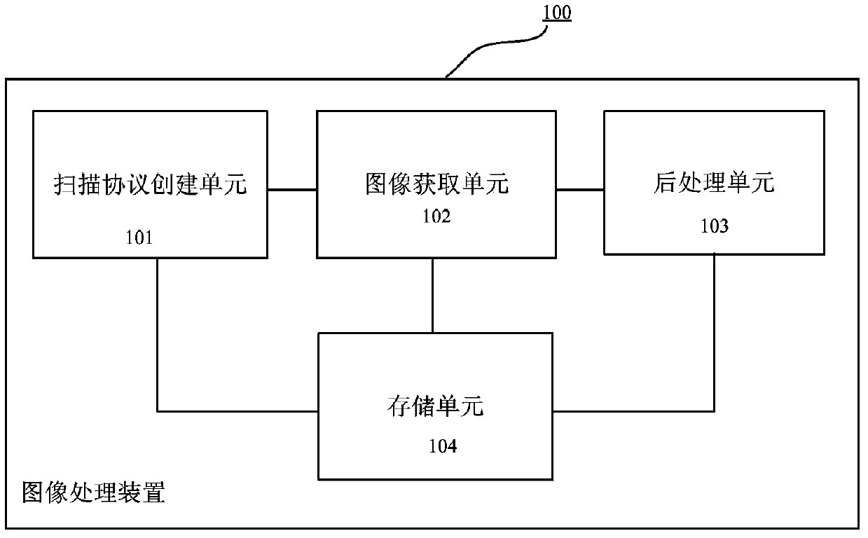
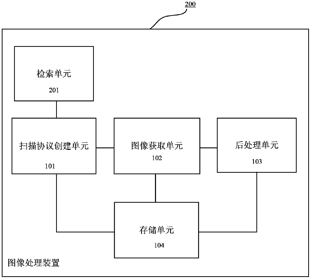
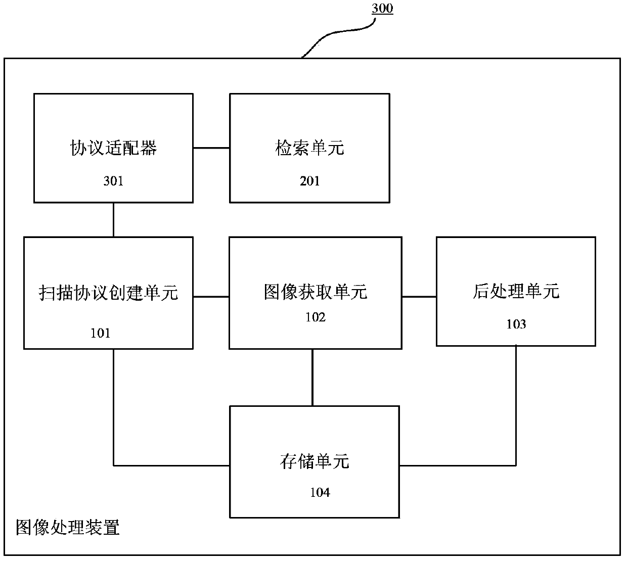
[0024] Such as figure 1 As shown, a structural block diagram of an image processing apparatus 100 according to an embodiment of the present application is shown. The image processing apparatus 100 includes: a scanning protocol creation unit 101 configured to create a scanning protocol for a specific part of the body of an object currently to be scanned; an image acquisition unit 102 configured to scan the object using an imaging device according t...


PUM


Abstract

An image processing apparatus according to an embodiment includes a scanning protocol creation unit, an image acquisition unit, a post-processing unit, and a storage unit. The scanning protocol creation unit creates a scanning protocol for a particular part of body of an object. The image acquisition unit acquires source image data by scanning the particular part using an imaging device according to scanning parameters in the scanning protocol. The post-processing unit performs post-processing on the source image data according to post-processing parameters in the scanning protocol to obtain processed image data. The storage unit stores the scanning protocol, or store the source image data and the processed image data or index identifiers thereof in association with parameters related to the scanning protocol as single job data of the object. The scanning protocol includes the scanning and the post-processing parameters, and information related to the imaging device and the post-processing unit.

Description

technical field [0001] The present application relates to the field of image processing, and in particular, relates to an image processing device and an image processing method for processing medical images, as well as medical image equipment. Background technique [0002] In the acquisition and / or processing of medical images, many parameters may need to be set, for example, when using magnetic resonance imaging (MRI), parameters such as TR (repetition time), TE (echo time), FOV (field of view) need to be set , when processing the obtained image, parameters such as motion correction, distortion correction, FA (flip angle) threshold, seed point, etc. need to be set. The setting of these parameters is crucial to the quality of the final image, and the consistency of parameter setting also helps to enhance the comparability between images. [0003] However, optimizing these parameters is a difficult and time-consuming task. Therefore, it is desirable to have a mechanism that...

Claims


Application Information

Patent Timeline
Patent Type & Authority Applications(China)
IPC IPC(8): G06T1/00G16H30/20
CPCG06T7/0012G06T2207/30004A61B6/032A61B6/488A61B6/5235A61B8/54A61B6/545G16H30/20
Inventor 许奇齐王晓婧张利军杉山敦子
Owner TOSHIBA MEDICAL SYST CORP
Learn More