Segmentation method and device for heart three-dimensional image

A three-dimensional image and heart technology, applied in the field of image processing, can solve the problems of inability to quickly realize image segmentation, increase the amount of calculation and complexity of segmentation methods, etc., reduce the amount of calculation and complexity, reduce the impact of local noise, and realize image segmentation. split effect

Active Publication Date: 2014-12-10
NEUSOFT MEDICAL SYST CO LTD
View PDF6 Cites 15 Cited by
  • Summary
  • Abstract
  • Description
  • Claims
  • Application Information

AI Technical Summary

Problems solved by technology

However, this segmentation method relies on the establishment of the initial three-dimensional model of the heart, and the process of establishing the model must undergo repeated training, which will increase the calculation and complexity of the segmentation method, and cannot quickly achieve image segmentation

Method used


Image

  • Segmentation method and device for heart three-dimensional image
  • Segmentation method and device for heart three-dimensional image
  • Segmentation method and device for heart three-dimensional image

Examples


Embodiment Construction

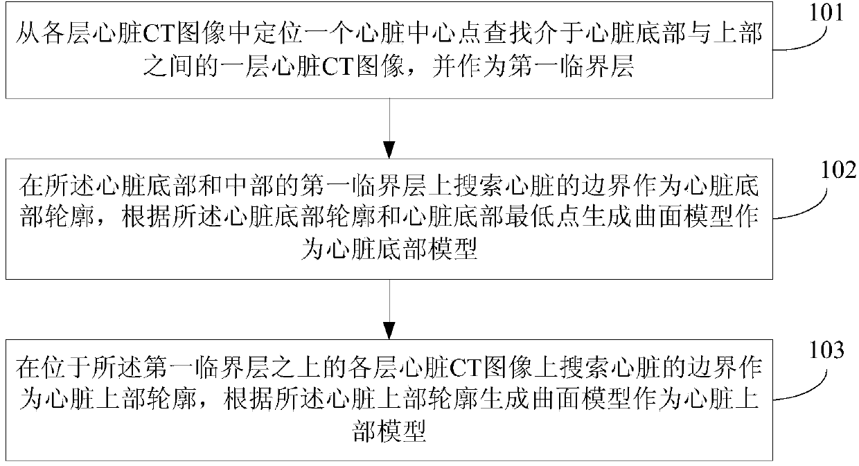
[0081] Embodiments of the present invention provide a method and device for segmenting a three-dimensional image of a heart. Considering that the heart has relatively complex morphological characteristics, that is, there is a relatively obvious boundary between the top of the heart and the surrounding adjacent tissues, while the bottom of the heart is connected to the diaphragm. Therefore, in the embodiment of the present invention, the critical layer is located first, that is, the critical layer between the top of the heart and the upper part of the heart, and the heart is divided into two parts, the upper part and the bottom part, according to this critical layer, and then each part of the heart is established segmentally. Finally, a complete three-dimensional heart model is formed by the upper heart model and the bottom heart model, and the three-dimensional image of the heart can be clearly displayed through the three-dimensional heart model.

[0082] In order to make the ...


PUM


Abstract

The invention discloses a segmentation method and device for a heart image. The method includes that a layer of heart computed tomography (CT) image between the bottom and the upper part of a heart is searched among all layers of scanned heart electronic computer X-ray tomography images to serve as a first critical layer; the boundary of the heart is searched on the first critical layer to serve as a heart bottom outline, and a surface model is generated according to the heart bottom outline and a heart bottom lowest point to serve as a heart bottom model; the boundary of the heart is searched on all layers of heart CT images above the first critical layer to serve as a heart upper part outline, and a surface model is generated according to the heart upper part outline to serve as a heart upper part model. According to the segmentation method and device for the heart three-dimensional image, the segmentation accuracy can be guaranteed, segmentation calculating amount and complexity can be reduced as far as possible, and image segmentation can be rapidly achieved.

Description

technical field [0001] The invention relates to the technical field of image processing, in particular to a method and device for segmenting a three-dimensional heart image. Background technique [0002] According to statistics, coronary heart disease (full name: coronary atherosclerotic heart disease) is one of the main diseases that seriously affect human health, and the incidence rate has been increasing year by year in recent years. Therefore, early detection and diagnosis of coronary heart disease, early treatment is particularly important. In the early detection of coronary heart disease, it mainly depends on the three-dimensional image of the patient's heart. Therefore, how to obtain a three-dimensional image of the patient's heart is the key to early detection of coronary heart disease. [0003] The heart is the power source of the human circulatory system and one of the most important organs in the human body. Since the heart is located in the chest cavity, above...

Claims


Application Information

Patent Timeline
Patent Type & Authority Applications(China)
IPC IPC(8): G06T7/00G06T17/00
CPCA61B6/032A61B6/503A61B6/5211G06T7/12G06T7/149G06T7/174G06T2207/10081G06T2207/20116G06T2207/30048G16H50/20
Inventor 李丙生何薇
Owner NEUSOFT MEDICAL SYST CO LTD
Why Eureka
Try Eureka