Method for segmenting and denoising lung parenchyma through lateral scanning and four-corner rotary scanning

A left and right scanning, lung parenchyma technology, applied in the field of lung parenchyma denoising, can solve the problem of not being able to segment and denoise various parts of the lung

Active Publication Date: 2015-01-07
TAIYUAN UNIV OF TECH
View PDF5 Cites 10 Cited by
  • Summary
  • Abstract
  • Description
  • Claims
  • Application Information

AI Technical Summary

Problems solved by technology

[0002] The shape of different parts of the human lung is different. It is mainly divided into three typical parts: upper, middle and lower. At present, the existing methods for lung parenchyma segmentation are mainly aimed at the middle part of the human lung, and cannot perform good segmentation on each part of the lung. denoising

Method used


Image

  • Method for segmenting and denoising lung parenchyma through lateral scanning and four-corner rotary scanning
  • Method for segmenting and denoising lung parenchyma through lateral scanning and four-corner rotary scanning
  • Method for segmenting and denoising lung parenchyma through lateral scanning and four-corner rotary scanning

Examples


Embodiment Construction

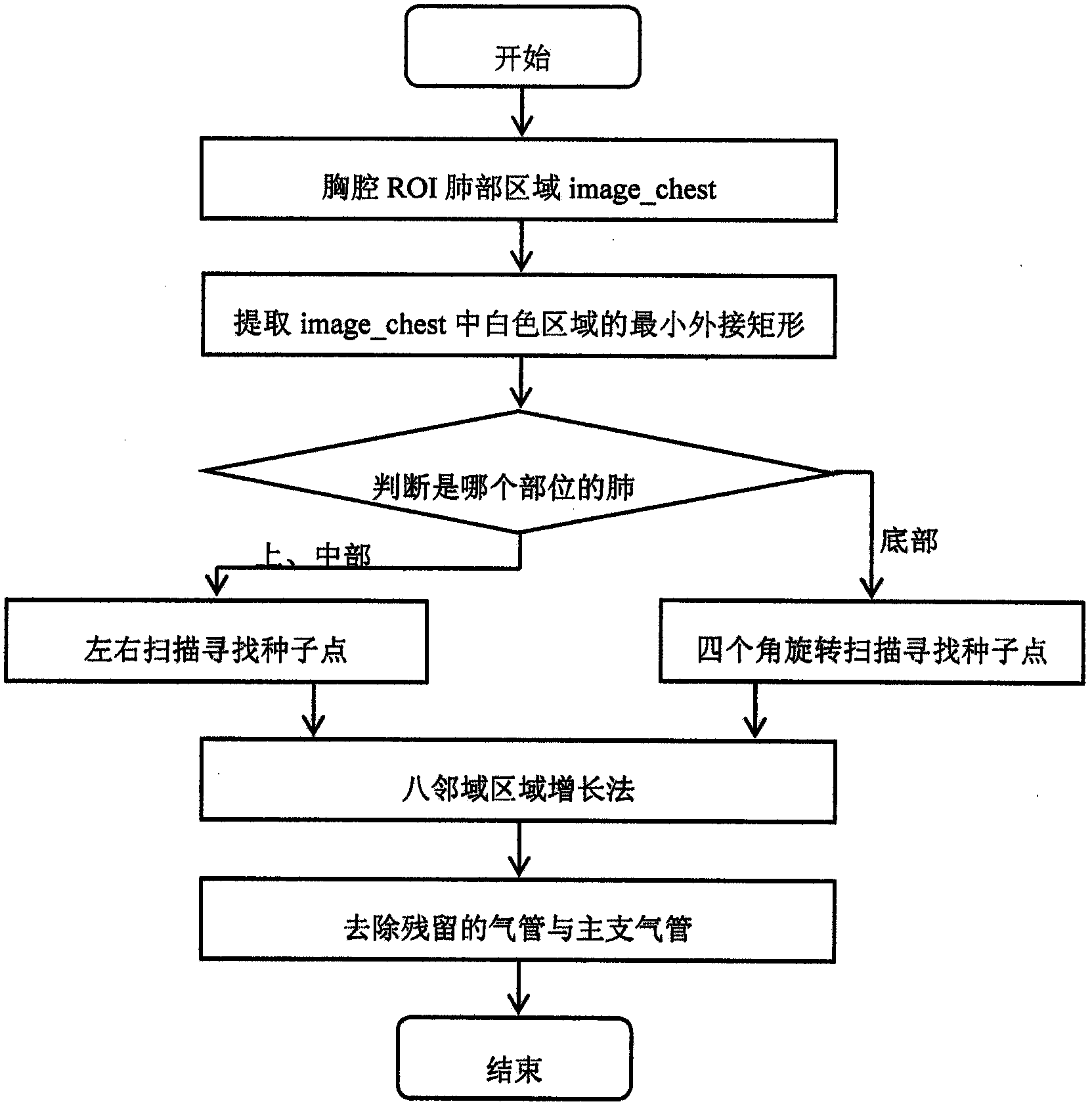
[0080] The present invention will be described in detail below in conjunction with specific embodiments.

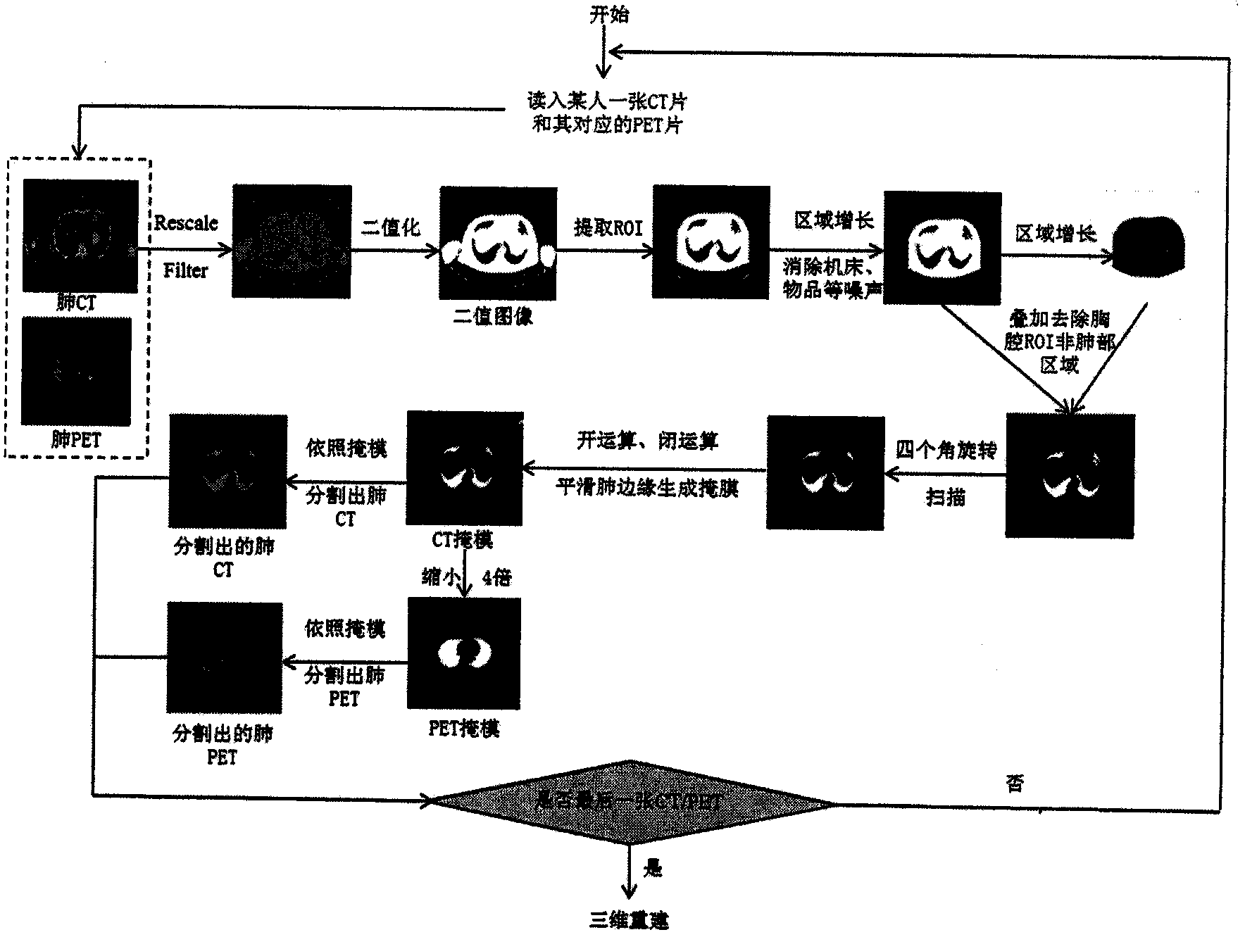
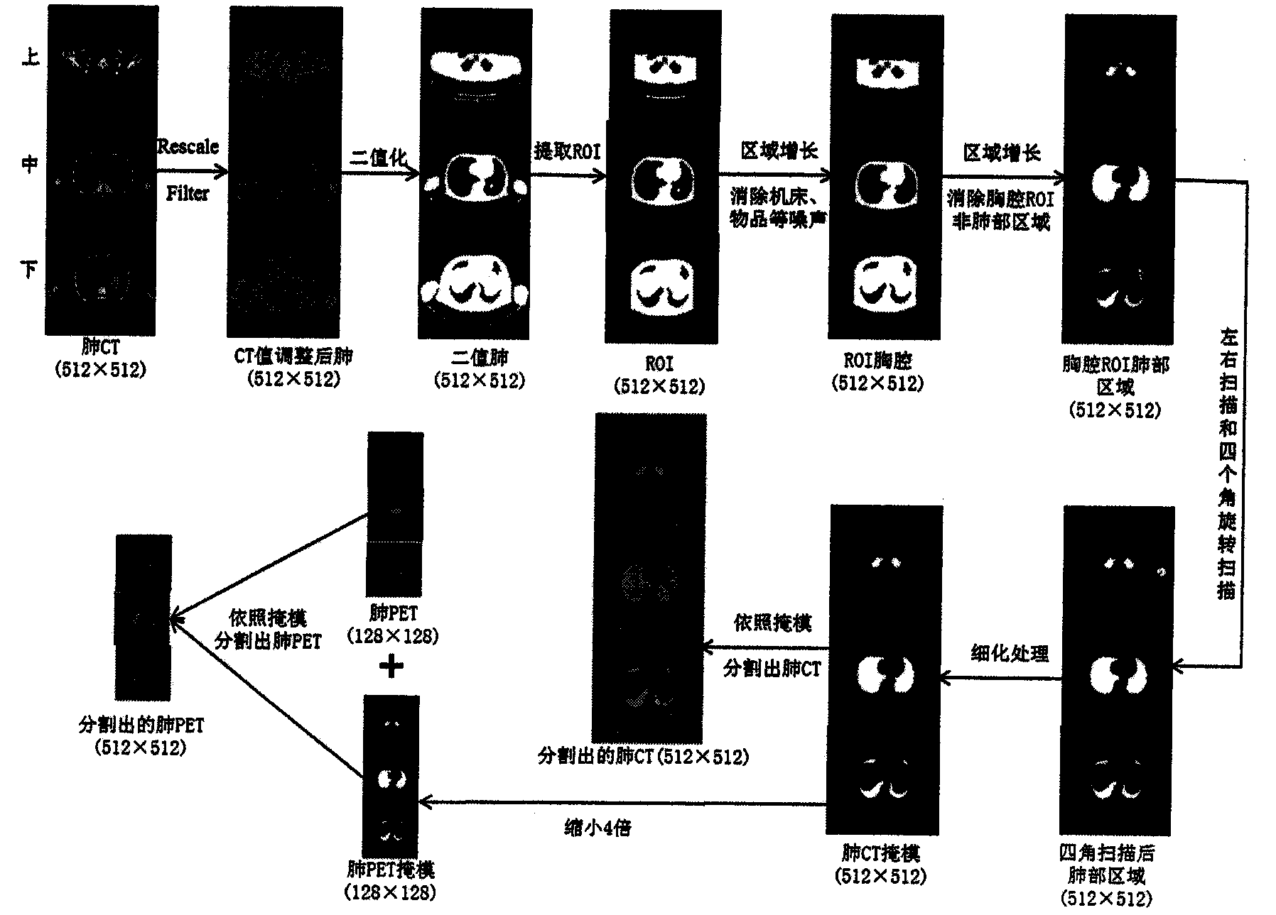
[0081] refer to figure 1 , 2 , the implementation process of the inventive method is as follows:

[0082] (1) Read the folder where someone's CT image sequence and its corresponding PET image sequence are located.

[0083] (2) Read a CT image of the person and its corresponding PET image from the folder read in in (1).

[0084] (3) Adjust the CT value between 0 and 255 through the RescaleIntensityImageFilter (image brightness adjustment filter) for the read-in CT image.

[0085] (4) The CT image after the CT value adjustment was binarized by threshold method based on iterative calculation to obtain a lung CT binary image.

[0086] (5) Extract the ROI (Region of Interest) from the lung CT binary image.

[0087] (6) The area growth method is adopted for the ROI to eliminate the noise of the machine tools and objects, and the chest CT binary image of the ROI is obtained...


PUM


Abstract

The invention discloses a method for segmenting and denoising the lung parenchyma through lateral scanning and four-corner rotary scanning. The method includes the steps that firstly, a minimum enclosing rectangle of an ROI lung region of the thoracic cavity is extracted; the minimum enclosing rectangle belonging to which portion of each lung is judged, lateral scanning or four-corner rotary scanning is selected according to the portion to which the minimum enclosing rectangle belongs, seed points are acquired, only lateral scanning is adopted in the upper portion of each lung, either lateral scanning or four-corner rotary scanning is adopted in the middle of each lung, and only four-corner rotary scanning is adopted in the bottom of each lung; eventually, residual trachea noise, main bronchus noise and other noise are removed according to an eight-neighborhood region growing method. The method is simple and easy to achieve, the segmentation speed is very high, for each image in a CT image sequence, a lung region of each image in a PET-CT image sequence can be completely, automatically and accurately segmented according to the method combining an iteration threshold value and morphology.

Description

technical field [0001] The invention relates to lung parenchyma denoising, in particular to a method for segmenting and denoising lung parenchyma by using left and right scans and four-angle rotation scans. Background technique [0002] The shape of different parts of the human lung is different. It is mainly divided into three typical parts: upper, middle and lower. At present, the existing methods for lung parenchyma segmentation are mainly aimed at the middle part of the human lung, and cannot perform good segmentation on each part of the lung. denoising. Sequence segmentation of lung images is the basis of 3D visualization of lungs. If images of various parts of lungs cannot be segmented correctly, the effect of 3D reconstruction of lungs will inevitably be affected. Contents of the invention [0003] The purpose of the present invention is to overcome the defects in the prior art and provide a simple, easy-to-implement, and very fast segmentation method for segmentin...

Claims


Application Information

Patent Timeline
Patent Type & Authority Applications(China)
IPC IPC(8): G06T7/00G06T5/00
CPCG06T5/002G06T7/12G06T2207/10081G06T2207/10104G06T2207/30061
Inventor 赵涓涓纪国华强彦胡洋洋廖晓磊吕鲤志
Owner TAIYUAN UNIV OF TECH
Why Eureka
Try Eureka