
Method and apparatus for single-particle localization using wavelet analysis

A particle, peacekeeping technology, applied in the direction of analyzing materials, image analysis, measuring devices, etc., can solve problems such as reducing calculation time

Active Publication Date: 2015-01-14
CENT NAT DE LA RECHERCHE SCI +1
View PDF2 Cites 23 Cited by
  • Summary
  • Abstract
  • Description
  • Claims
  • Application Information

AI Technical Summary

Problems solved by technology

This scheme greatly reduces computing time, but requires the use of dedicated graphics processing unit (GPU) hardware architecture

Method used


Image

  • Method and apparatus for single-particle localization using wavelet analysis
  • Method and apparatus for single-particle localization using wavelet analysis
  • Method and apparatus for single-particle localization using wavelet analysis

Examples


Embodiment Construction

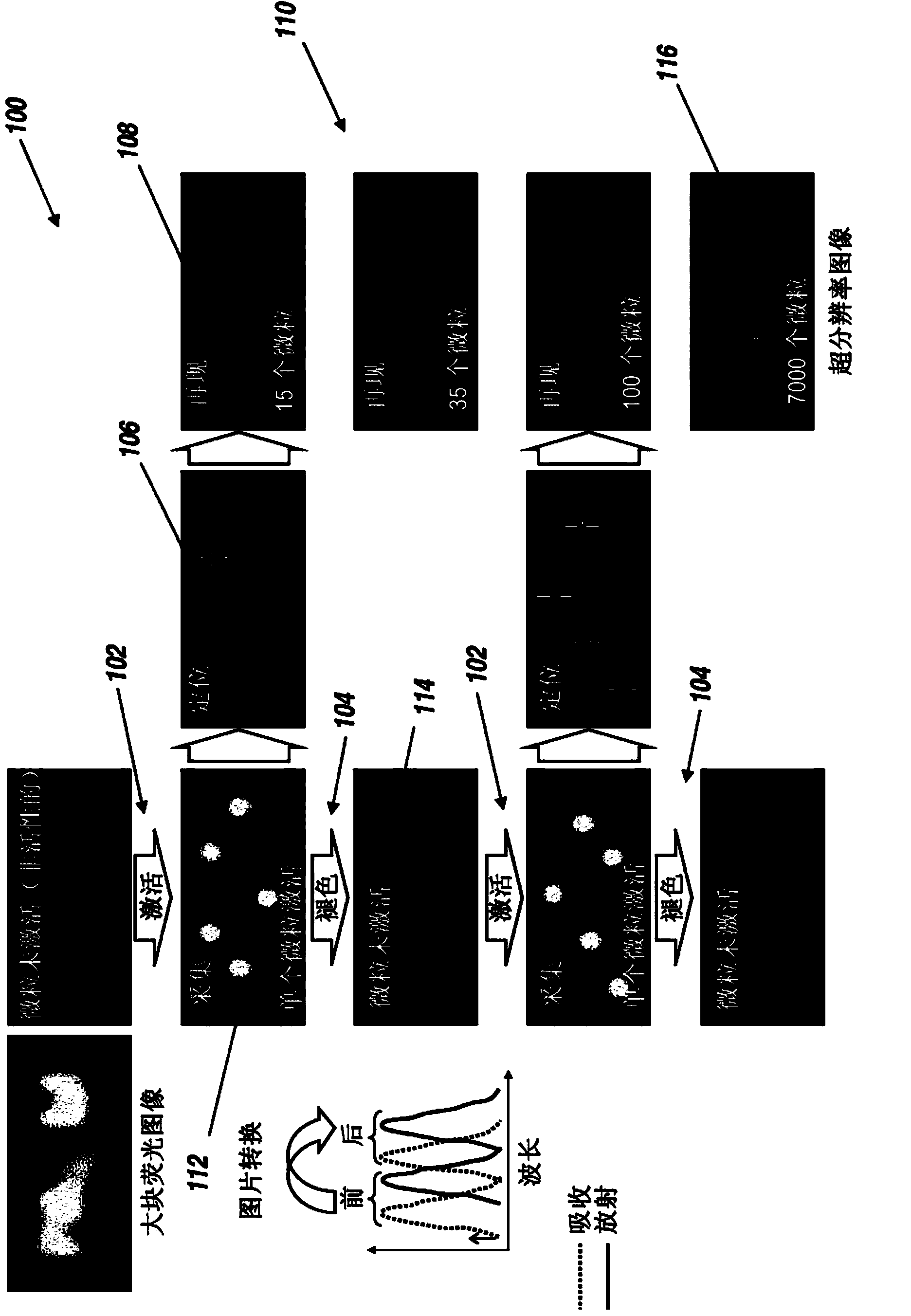
[0036] Wavelet segmentation for particle detection and centroid localization addresses limitations due to time-consuming data analysis. The devices and techniques of the present invention can be used for single-particle imaging, super-resolution imaging, particle tracking, and / or to manipulate fluorescent particles, including but not limited to biological cells, molecules (e.g., fluorescent proteins), organic fluorophores, Quantum dots, carbon nanotubes, diamond, metal or dielectric beads, and particles labeled with fluorophores. There are at least two advantages to this wavelet method: its processing time is more than an order of magnitude faster than methods involving two-dimensional (2D) Gaussian fitting; and its good localization accuracy can approach nanometer scale. In addition, 2D wavelet localization can be used with additional fitting techniques to provide real-time or near real-time three-dimensional (3D) localization.

[0037] Single-particle, wavelet-based localiz...


PUM


Abstract

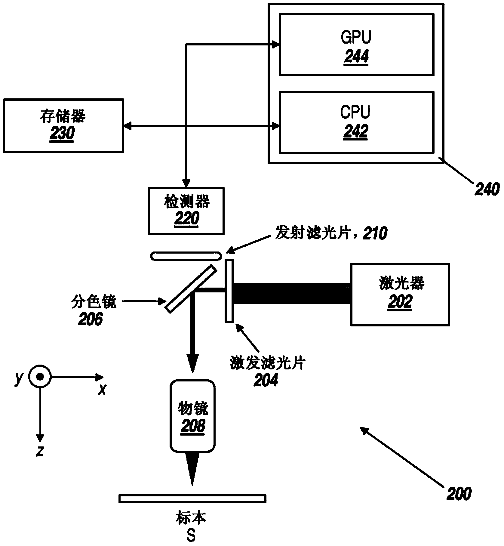
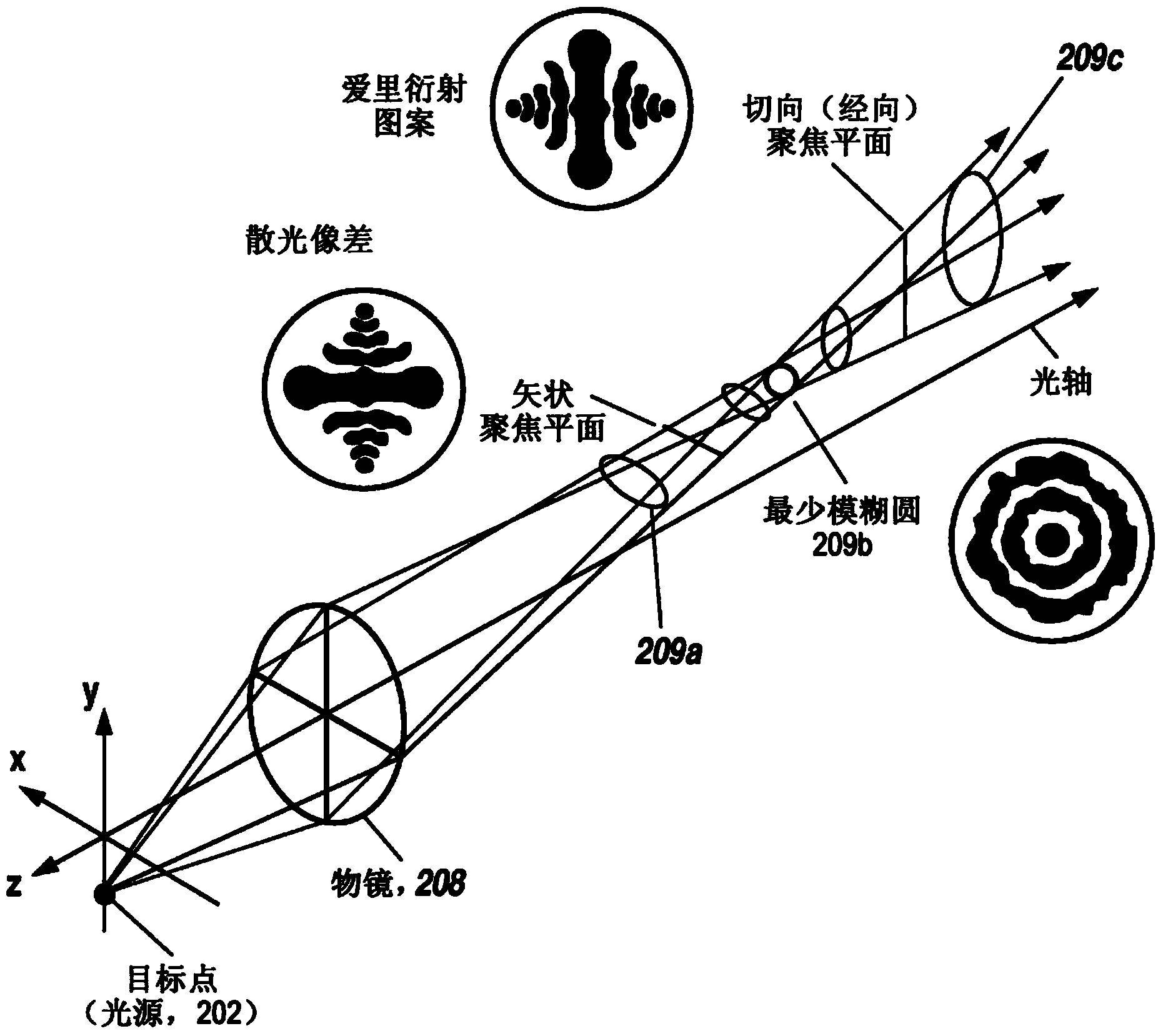
Accurate localization of isolated particles is important in single particle based super-resolution microscopy. It allows the imaging of biological samples with nanometer-scale resolution using a simple fluorescence microscopy setup. Nevertheless, conventional techniques for localizing single particles can take minutes to hours of computation time because they require up to a million localizations to form an image. In contrast, the present particle localization techniques use wavelet-based image decomposition and image segmentation to achieve nanometer-scale resolution in two dimensions within seconds to minutes. This two-dimensional localization can be augmented with localization in a third dimension based on a fit to the imaging system's point-spread function (PSF), which may be asymmetric along the optical axis. For an astigmatic imaging system, the PSF is an ellipse whose eccentricity and orientation varies along the optical axis. When implemented with a mix of CPU / GPU processing, the present techniques are fast enough to localize single particles while imaging (in real-time).

Description

Background technique [0001] The field of optical microscopy for biological applications has made a quantum leap as the technology has advanced to single particle detection. In recent years, single particle experiments using imaging techniques in biology and biophysics have become routine in many laboratories, providing new insights into numerous biological processes. In most cases, the first step in the quantitative analysis of single particle experiments is to determine the position of the particle with sub-pixel precision in the nanoscale range (just below the diffraction limit of light microscopy). For example, the precise position of fluorescently labeled proteins in sequential time-lapse images can be used to determine the diffusion properties of thin-film proteins or to unravel the stepping mechanisms of molecular motors. [0002] In recent years, several super-resolution optical microscopy techniques have been developed that go beyond the diffraction limit of light in ...

Claims


Application Information

Patent Timeline
IPC IPC(8): G02B21/00G01N21/64G06T7/00
CPCG06T7/0081G06T7/0042G02B21/0076G02B21/365G06T7/004G01N21/6458G06T2207/20064G06T7/0095G06T7/11G06T7/168G06T7/73G06T7/70G01N21/64G01N2015/1486G01N2015/0238G06V20/69G06V20/695G06F2218/06G06F2218/10G06T2207/10056G06T2207/10064
Inventor 让-巴蒂斯·西巴里塔
Owner CENT NAT DE LA RECHERCHE SCI
Learn More