Supercharge Your Innovation With Domain-Expert AI Agents!

Medical image processing apparatus and medical image processing method

A medical image and processing device technology, applied in image data processing, image enhancement, image generation, etc., can solve problems such as narrow calcification features or other state assessments

Active Publication Date: 2015-04-15
TOSHIBA MEDICAL SYST CORP
View PDF4 Cites 4 Cited by
  • Summary
  • Abstract
  • Description
  • Claims
  • Application Information

AI Technical Summary

Problems solved by technology

The presence of such calcified features may make it difficult or impossible to assess for stenosis or other conditions

Method used


Image

  • Medical image processing apparatus and medical image processing method
  • Medical image processing apparatus and medical image processing method
  • Medical image processing apparatus and medical image processing method

Examples


Embodiment Construction

[0019] Hereinafter, a medical image processing device and a medical image processing method according to the present embodiment will be described with reference to the drawings. In addition, in the following description, the same code|symbol is attached|subjected to the component which has substantially the same function and structure, and repeated description is performed only when necessary.

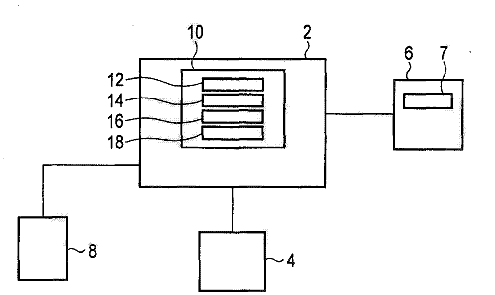
[0020] image 3 It is a diagram showing an example of a block configuration of a medical image processing system according to an embodiment. Such as image 3 As shown, the medical image processing device 2 according to one embodiment is, for example, a personal computer (PC) or a workstation. To the medical image processing apparatus 2, a display device 4, a scanner 6, a user input device 8, and the like are connected. There can be one or more user input devices 8 . At this point, connect the computer keyboard and mouse.

[0021] The scanner 6 is any suitable type of CT scanner ca...


PUM


Abstract

Apparatus for performing an imaging procedure comprising processing volumetric image data comprising an image processing unit configured to obtain first image data representative of a region including at least one vessel and at least one associated feature, and second image data representative of at least part of the region, an image refinement unit configured to process the first image data and the second image data to produce a combined image representative of the at least one vessel, wherein the associated feature is removed or reduced in intensity in the combined image, and a rendering unit configured to render the combined image as a simulated view that simulates a view obtainable from an alternative imaging procedure.

Description

technical field [0001] This embodiment relates to a medical image processing device and a medical image processing method. Background technique [0002] Coronary artery stenosis is an obvious indicator of coronary artery disease, and various imaging techniques and diagnostic techniques have been developed in order to identify and visualize the site including the stenosis. [0003] Catheter coronary angiography has traditionally been considered the most accurate imaging technique for the assessment of coronary artery stenosis. Catheter coronary angiography, however, is an invasive procedure requiring positioning of the catheter relative to each coronary ostium, causing discomfort to the patient and possibly some danger. In addition, this technique uses X-ray fluoroscopy with exposure of the patient to high radiation doses of radiation. figure 1 is a diagram showing an example of a 2D image obtained using a known catheter coronary angiography technique. [0004] In addition...

Claims


Application Information

Patent Timeline
IPC IPC(8): G06T11/00A61B6/03
CPCG06T2207/10081G06T2207/30101G06T2210/41G09G5/377G06T15/08G06T7/0012G06T7/30G06T2207/20016G06T2207/20224G06T2207/30048G09G2320/066G09G2380/08
Inventor M·拉择图A·埃金耶米M·H·巴哈蒂J·巴纳比S·梅勒
Owner TOSHIBA MEDICAL SYST CORP
Features
  • R&D
  • Intellectual Property
  • Life Sciences
  • Materials
  • Tech Scout
  • Unparalleled Data Quality
  • Higher Quality Content
  • 60% Fewer Hallucinations
Learn More