
Adaptive Enhancement Method Based on Mammogram

An image enhancement and line image technology, applied in the field of medical image processing, can solve problems such as reducing the signal-to-noise ratio, and achieve the effects of suppressing noise, increasing image contrast, and improving overall contrast.

Inactive Publication Date: 2018-01-19
BEIJING UNIV OF TECH
View PDF2 Cites 0 Cited by
  • Summary
  • Abstract
  • Description
  • Claims
  • Application Information

AI Technical Summary

Problems solved by technology

Although the above method has achieved a good enhancement effect to a certain extent, it also enhances the noise in the image while enhancing the image, which reduces the signal-to-noise ratio, so there are still certain limitations.

Method used


Image

  • Adaptive Enhancement Method Based on Mammogram
  • Adaptive Enhancement Method Based on Mammogram
  • Adaptive Enhancement Method Based on Mammogram

Examples


Embodiment Construction

[0037] The extraction process is described in detail with reference to the accompanying drawings and practical examples. The image data used came from mammography images in the MIAS database. The size of each mammogram image is 1024*1024 pixels.

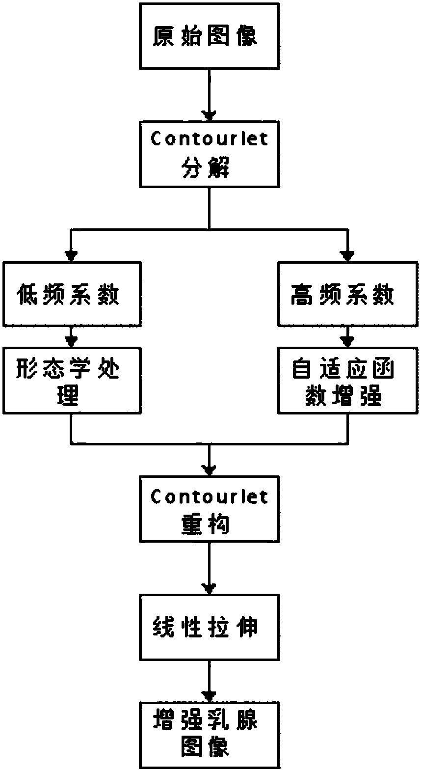
[0038] The flow chart of the self-adaptive enhancement method based on mammogram of the present invention is as follows figure 1 shown, including the following steps:

[0039] Step 1, perform non-sampled contourlet wave 3-layer decomposition on the input breast image, so as to decompose the breast image into low-frequency coefficient sub-image X and high-frequency coefficient sub-image in different directions Where j represents the number of decomposed layers j=1,2,3. k represents different decomposition directions.

[0040] In step 2, the morphological method is used to process the low-frequency coefficient sub-image X obtained through contourlet transformation.

[0041] 2.1. Use the Top-hat transformation to process the ima...


PUM


Abstract

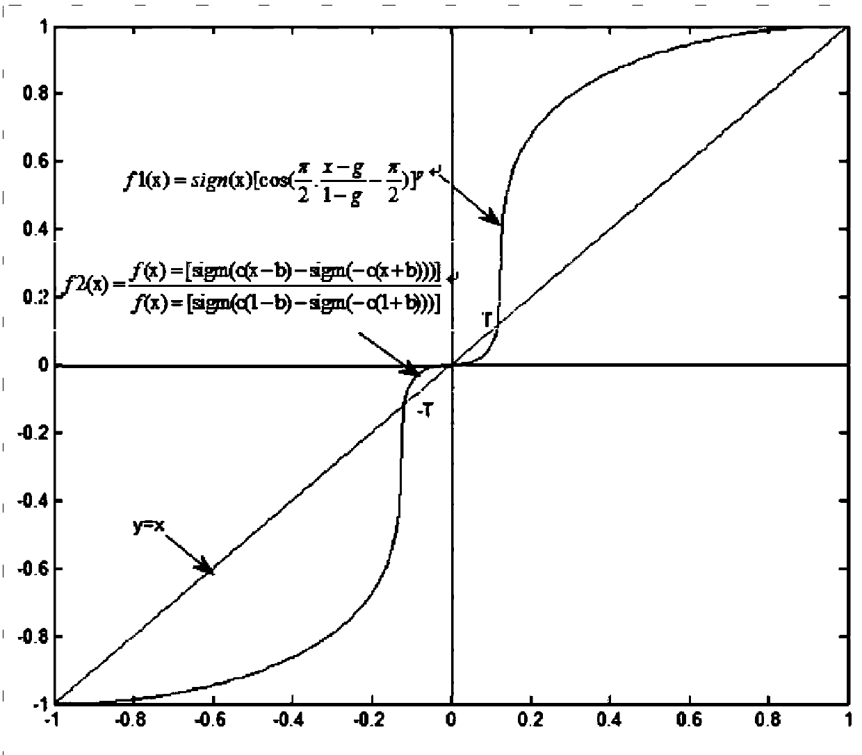
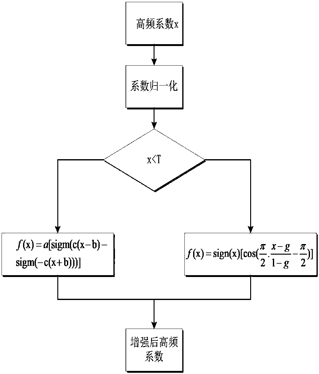
The invention discloses an adaptive enhancement method based on a mammogram. The image is decomposed by contourlet transformation to obtain high-frequency sub-images and a low-frequency sub-image in different directions. Use morphology to process low-frequency subgraph coefficients. The high-frequency sub-image coefficients in different directions are enhanced by using the constructed adaptive enhancement function. Perform contourlet reconstruction on the processed high-frequency coefficient sub-image and low-frequency coefficient sub-image to obtain an enhanced mammogram image. The invention can enhance the weak edges and calcified points in the image while suppressing the image noise, and effectively improve the image contrast. The enhancement effect is better than the existing unsharp masking method and adaptive histogram equalization method.

Description

technical field [0001] The invention relates to the field of medical image processing, in particular to an adaptive enhancement method based on mammograms. Background technique [0002] Breast cancer is one of the most common malignant tumors in women with high morbidity and mortality. According to the statistics of the World Health Organization, there are 1.2 million new cases of breast cancer every year in the world, and the death toll is 500,000. In addition, the incidence of breast cancer is still on the rise, which makes the prevention and treatment of breast cancer more and more people's attention. [0003] In recent years, with the development of modern medical imaging technology, more and more detection methods have been applied to the early detection of breast cancer. Mammography has become a reliable method for early detection of breast cancer due to its low price, simple operation, and high spatial resolution. However, doctors also face many difficulties when u...

Claims


Application Information

Patent Timeline
Patent Type & Authority Patents(China)
IPC IPC(8): G06T5/00A61B6/00
Inventor 吴水才彭庆涛高宏建吴薇薇
Owner BEIJING UNIV OF TECH
Learn More