

Method for determining biology activity of PD-1 pathway inhibitor
A biologically active, PD-1 technology, applied in biochemical equipment and methods, microbial assay/inspection, cells modified by the introduction of foreign genetic material, etc., can solve the problem of long time, high cost, and large method variability. question
- Summary
- Abstract
- Description
- Claims
- Application Information
AI Technical Summary
Problems solved by technology
Method used
Image

Examples
Embodiment 1
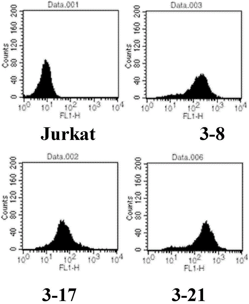
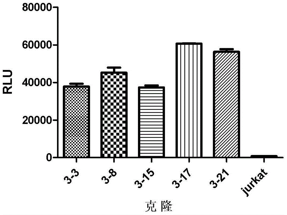
[0086] Example 1: Preparation of effector cells expressing PD-1 and NFAT-luciferase
[0087] First, pcDNA3.1(+) / PD-1 (Beijing Sino Biological Technology Co., Ltd.) and pGL4.30[Luc2P / NFAT-RE] (Promega (Beijing) Biotechnology Co., Ltd. company) plasmids were co-transfected into jurkat cells (ATCC), and then cultured in a medium containing 1 μg / ml puromycin and 1 mg / ml geneticin to obtain effector cells expressing PD-1 and NFAT-luciferase . Using jurkat cells as a control, the expression of PD-1 in the obtained cells was analyzed by flow cytometry (FACs), and the results are shown in figure 1 . Stimulate with mouse anti-human CD3 antibody (BD Biosciences, cat.555336), by Bright-Glo TM Kit (Promega, cat.E2610) was used to measure the expression of luciferase (luciferase), and the results of luciferase activity were found in figure 2. Thus, the effector cells of the present invention are obtained. The stable cell line JurKat / PD-1 / NFAT was obtained by selecting the clone wit...
Embodiment 2
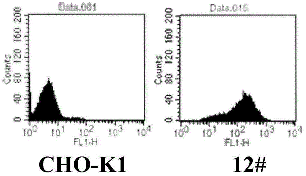
[0088] Example 2: Preparation of target cells expressing αCD3TM and PD-L1
[0089] The pcDNA3.1(+) / αCD3TM and pcDNA3.1(+) / PD-L1 plasmids (Beijing Sino Biological Technology Co., Ltd.) were co-transfected into CHO-K1 cells ( ATCC), the transfected cells were then cultured in medium containing 1 μg / ml puromycin and 200 μg / ml hygromycin B. Using CHO-K1 as a control, the expression levels of αCD3TM and PD-L1 in the resulting cells were detected using donkey anti-mouse antibody and PD-L1 antibody. For the results of FACs of αCD3TM and PD-L1 expression image 3 and Figure 4 . Thus, the target cells of the present invention are obtained. The clone with higher expression level was selected to obtain a stable cell line CHO-K1 / αCD3TM / PD-L1, which was deposited in China Microorganism Culture Collection Management Committee General Microbiology on December 30, 2014 with the registration number CGMCC No.10299. center.
Embodiment 3
[0090] Example 3: Determination of biological activity of PD-1 monoclonal antibody and PD-L1 monoclonal antibody according to the method of the present invention
[0091] (1) Prepare CHO-K1 / αCD3TM / PD-L1 target cell suspension, use DMEM / F12+10% FBS medium according to 2.5×10 4 cells / well, spread evenly in 96-well plate at a density of 100 μl / well (corning), 37°C, 5% CO 2 Incubate overnight in the incubator;
[0092] (2) Use 1640+10% medium to prepare different concentrations of PD-1 monoclonal antibody (Nivolumab) or PD-L1 monoclonal antibody (MPDL3280A) solutions and dilute to 10 μg / mL, then down 3-fold ratio dilution 7 At each concentration point, absorb the DMEM / F12+10% FBS medium in the above-mentioned 96-well plate, and add the prepared monoclonal antibody solution of PD-1 monoclonal antibody or PD-L1 monoclonal antibody to 50 μl / well In 96-well plate;
[0093] (3) Use 1640+10% medium to prepare JurKat / PD-1 / NFAT effector cell suspension, and follow the 5×10 4 per well,...
PUM
 Abstract
Description
Claims
Application Information