

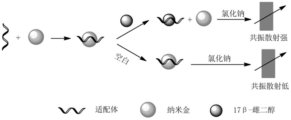
Method for detecting 17bata-estradiol by using resonance scattering spectrometry based on functional nucleic acids
A resonance scattering, functional nucleic acid technology, applied in fluorescence/phosphorescence, material excitation analysis, etc., can solve the problems of prone to false positives, long detection time, high cost, and achieve the effect of significant technological progress, low cost, and good stability.
- Summary
- Abstract
- Description
- Claims
- Application Information
AI Technical Summary
Problems solved by technology
Method used
Image

Examples
Embodiment 1
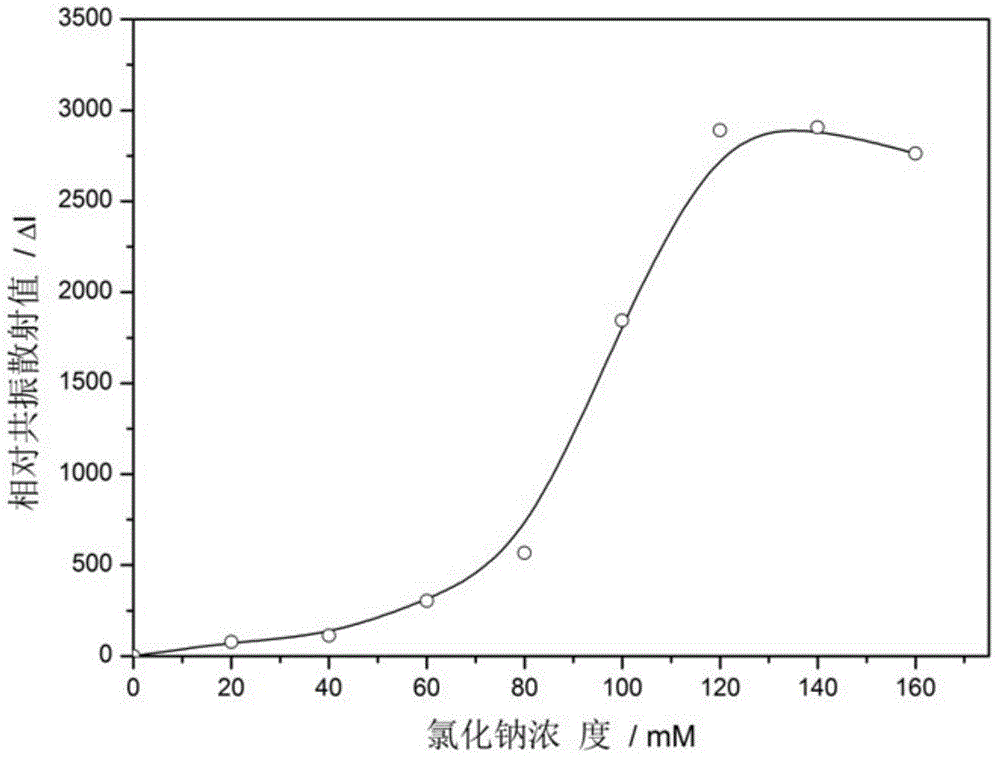
[0028] Example 1 Sodium chloride concentration in the optimized system
[0029] In a 1.5mL centrifuge tube, add 100μL of nano-gold solution, then add propanesulfonic acid buffer (concentration is 10mmol / L, pH is 7.0), mix well, and sequentially add sodium chloride solution with a concentration range of 0-160mmol / L, After five minutes, the resonance scattering value was measured, the relative resonance scattering value was obtained, and a curve was drawn. The sodium chloride concentration corresponding to the highest peak was 120 mmol / L, such as figure 2 shown.
Embodiment 2
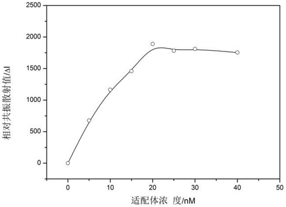
[0030] Example 2 Optimizing the concentration of 17β-estradiol aptamer in the system
[0031] The 17β-estradiol aptamer was purchased from Shanghai Shenggong. The received sample was prepared into a 1 μmol / L mother solution, and then the aptamer with a concentration range of 0-40 nmol / L was added to 100 μL of nano-gold solution. For aptamer-gold nanoparticles, after incubation for 15 minutes, add a certain concentration of estradiol solution, incubate for 15 minutes, add the optimized sodium chloride solution, adjust the volume to 500 μL with propanesulfonic acid buffer, and measure the resonance scattering value after five minutes , draw a curve, the aptamer concentration corresponding to the highest peak is 20nmol / L, such as image 3 shown.
Embodiment 3
[0032] Embodiment 3 The process of using the method of the present invention to detect whether the milk sample contains 17β-estradiol specifically includes the following steps:
[0033](1) Prepare a detection system with known 17β-estradiol concentration: take 11 centrifuge tubes to which 10 μL and 1 μmol / L 17β-estradiol aptamer were added respectively, add 100 μL nano-gold solution, mix well and place the centrifuge tubes into the centrifuge tubes. Incubate at 25°C for 15min, and then add 10μL of 17β-estradiol solutions of different concentrations respectively, so that the content of 17β-estradiol in the entire detection system is maintained at 0.1-3.0μmol / L, and then fully mixed before Incubate the centrifuge tube at 25°C for 15 min, add 30 μL, 2 mol / L sodium chloride solution to the above mixture, mix well, and adjust the buffer volume to 500 μL, which is reserved for the following determinations;
[0034] (2) Take another centrifuge tube and replace the 17β-estradiol solut...
PUM
| Property | Measurement | Unit |
|---|---|---|
| Concentration | aaaaa | aaaaa |
Abstract
Description
Claims
Application Information