

CT scan perfusion method and device
A technology of CT scanning and CT value, which is applied in computerized tomography scanners, echo tomography, etc., can solve the problems of poor compliance and hinder the practical application of CT perfusion technology, and achieve good compliance, simple and easy operation, and reduce radiation effect of dosage
- Summary
- Abstract
- Description
- Claims
- Application Information
AI Technical Summary
Problems solved by technology
Method used
Image

Examples
no. 1 approach
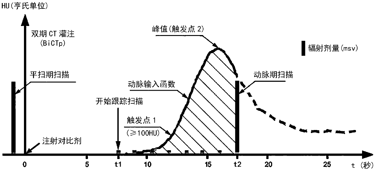
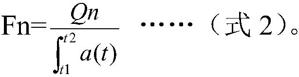
[0020] Next, in the CT scanning perfusion method of the present invention, in order to obtain the increase Q of the CT value in the organ caused by the contrast medium perfusion, before injecting the contrast medium, a CT plain scan is performed on the organ of the subject. In the description, this method is referred to as dual-phase (plain scan phase and arterial phase) CT perfusion, and this is taken as an example to describe the composition of the present invention in detail.
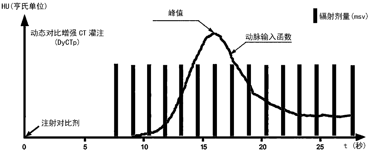
[0021] figure 1 It is a dynamic data scanning diagram showing an embodiment of dual-phase CT perfusion of the present invention. exist figure 1 In , the abscissa is the time axis, the point at which the contrast agent starts to be injected is set as time 0, and the ordinate represents the CT value (HU).
[0022] The specific implementation steps of the dual-phase CT perfusion performed on the subject are performed in the following order.
[0023] (flat scan)
[0024] Before injecting contrast med...
no. 2 approach
[0053] In the second embodiment, except that the steps of "scanning and tracking" and "arterial phase scanning" are slightly different from those of the first embodiment, the content of other steps is the same as that of the first embodiment, and their descriptions are omitted here.
[0054] The differences between the second embodiment and the first embodiment in the "scan tracking" step and the "arterial phase scan" step will be mainly described below.
[0055] In the "scanning and tracking" step of the second embodiment, during the scanning and tracking of the iodine contrast agent with a low radiation dose, the change of CT value with time is depicted as the time density curve of the artery (arterial input function), which is consistent with the first In one embodiment, after the CT value of the artery reaches a certain threshold (trigger point 1), a predetermined time is delayed, and then the arterial phase scanning of the conventional radiation dose is started on the arte...
no. 3 approach
[0057] The increase Q of the CT value, when used in dual-source CT or spectral CT, is obtained from the arterial phase scan through dual-energy subtraction or spectral subtraction.
[0058] The third embodiment is that when the CT scanning perfusion method of the present invention is used for dual-energy CT (dual-source CT) or energy spectral CT, the "scanning in the plain scan" is omitted, and the increase Q of the CT value is obtained by scanning the arterial phase. Except for the direct acquisition of dual-energy subtraction or spectral subtraction, it is the same as the first embodiment, and the description of the same parts will be omitted here.
PUM
 Abstract
Description
Claims
Application Information