Single molecular detection method of let-7a in human lung cells

A let-7a, single-molecule detection technology, applied in biochemical equipment and methods, microbial determination/inspection, etc., can solve problems such as failure to achieve quantitative detection of targets and no practical application value, and achieve great application potential, The effect of good specificity and sensitivity

Inactive Publication Date: 2015-07-08
SHANDONG UNIV
View PDF1 Cites 10 Cited by
  • Summary
  • Abstract
  • Description
  • Claims
  • Application Information

AI Technical Summary

Benefits of technology

This technology works well with small mature proteins called micro-ribonucleic acid or MNRs which play an important role during cellular growth processes such as differentiation, developmental regulation, cancer metastasis, stem cell renewing, etc., but they may have harmful side effect on other organisms if taken carelessly due to their ability to affect gene expression patterns. By analyzing these molecules from various sources like blood plasma, urine, saliva, sputum, tissue sections, brain cords, bone marrow aspirates, tumor specimens, skin swabs, hair follicles, cervix uteri, stomach lining fluid, liver fluids, chick embryos, hepatocytes, placentas, muscle spindlets, nerve fibers, cardiac trovesicles, kidneys, pancreases, adrenals, ovary glands, breast milk, pancreatitis, ovaries, testes, thymus primordium, heart disease patients' circulating DNA, and others weigh heavily towards understanding how this new discovery could help develop better diagnoses and treatments against diseased states caused by abnormal protein production.

Problems solved by technology

Technologies described in this patents involve developing various techniques called qSRP(quantum SR Probing) and SQDN (single nucleotide polymerase/DNA sequencing). These technics aim at identifying minute amounts of targets within cells without being affected by background noise sources like other proteins. They require precise measurements while avoiding any interference caused by unrelated substances such as protein interactions. Additionally, they rely heavily upon immune responses during inflammatory processes, leading to potential misidentifications even if some were present. To address these issues, we developed a novel technique called SSCRM (Single Structure Quantitation Microscopy)-based Methodology: The technical problem addressed in this study is improving the ability of detecting bioactive compounds specifically associated with certain types of diseases based on their unique characteristics.

Method used


Image

  • Single molecular detection method of let-7a in human lung cells
  • Single molecular detection method of let-7a in human lung cells
  • Single molecular detection method of let-7a in human lung cells

Examples


Embodiment 1

[0046] Example 1: Single molecule detection of target let-7a

[0047] Specific steps are as follows:

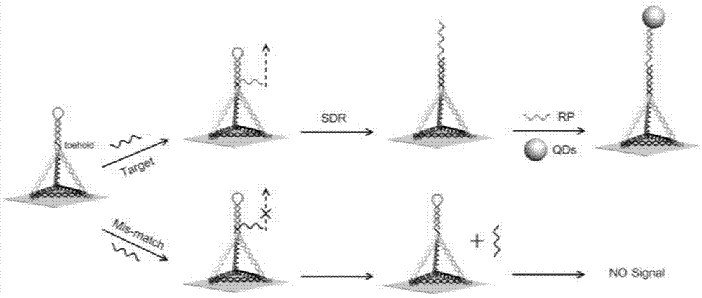
[0048] (1) DNA tetrahedron assembly:

[0049] Dilute the four DNA strands (A-HP, B-NH2, C-NH2 and D-NH2) to 50 μM respectively, take 2 μL each into 92 μl TM buffer, mix well, anneal at 95 ° C for 2 min, and place in ice bath to obtain DNA tetrahedron solution, wherein the concentration of the DNA tetrahedron is 1 μM.

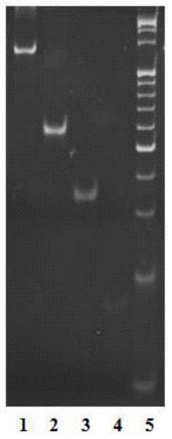
[0050] Electrophoresis verification: use 9.6% polyacrylamide gel electrophoresis to verify the DNA tetrahedron solution.

[0051] In order to characterize the assembly of the DNA tetrahedron, this embodiment carried out polyacrylamide gel electrophoresis verification, such as figure 2 , compared with other single-stranded structures and structures lacking one or two strands, the DNA tetrahedron moves slowly, which is consistent with previous reports and confirms the successful synthesis of the DNA tetrahedral structure. 36,37

[0052] (2) Construction of ...

Embodiment 2

[0057] Embodiment 2: the sensitivity investigation of method

[0058] Specific steps are as follows:

[0059] Same as steps (1) and (2) described in Example 1.

[0060] (3) Sensitivity investigation of miRNA detection

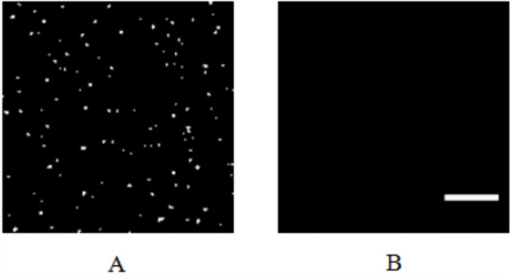
[0061]First, 50 μL of miRNA at different concentrations (1.0pM, 0.8pM, 0.5pM, 0.3pM, 0.1pM, 0.05pM and 0.005pM) were added to the prepared substrate, and incubated at 37°C for 2h. After washing with PBS-T and PBS buffer three times respectively, 50 μL of bio-RP (0.1 nM) was added and incubated at 37° C. for 2 h. After washing with PBS-T and PBS buffer three times, 50 μL of QDs (0.1 nM) was added and incubated at 37° C. for 2 h. After washing with PBS-T and PBS buffer three times, 50 μL of PBS buffer was added, imaging was performed under a fluorescence episcopic microscope, and miRNA was quantified by counting the number of fluorescent spots.

[0062] The present invention detects miRNAs of different concentrations in the buffer solution, such as Figure 4...

Embodiment 3

[0063] Embodiment 3: the specificity investigation of method

[0064] Specific steps are as follows:

[0065] Same as steps (1) and (2) described in Example 1.

[0066] (3) Specificity investigation of miRNA detection

[0067] First, 50 μL of different let-7 family member miRNAs were added to the prepared tetrahedral substrate, and incubated at 37°C for 2 hours. After washing with PBS-T and PBS buffer three times respectively, 50 μL of bio-RP (0.1 nM) was added and incubated at 37° C. for 2 h. Wash with PBS-T and PBS buffer three times respectively, add 50 μL QDs (0.1 nM), and incubate at 37° C. for 2 h. After washing with PBS-T and PBS buffer three times, 50 μL of PBS buffer was added, and imaging was performed under a fluorescence episcopic microscope. The specificity of the method was investigated by counting the number of fluorescent spots produced by different targets.

[0068] miRNAs are characterized by high sequence similarity among homogeneous groups, so it is a g...


PUM


Abstract

The invention discloses a single molecular detection method of let-7a in human lung cells. The single molecular detection method comprises the following steps: (1) assembling a DNA (Deoxyribose Nucleic Acid) tetrahedral probe to obtain a DNA tetrahedral solution; (2) preparing a silanization glass substrate, then adding the DNA tetrahedral solution to the substrate, and inoculating to obtain a DNA tetrahedral substrate; (3) carrying out single molecular detection on let-7a, namely, detecting by adopting a standard addition method, firstly respectively adding materials to be detected to a Tris-HCl solution of standard let-7a with stagewise concentration to prepare samples to be detected; then adding a biotinylated ssDNA probe and a streptavidin quantum dot, imaging by using a fluorescent shooting microscope, respectively checking the number of phosphor dots, making a standard curvilinear equation, and calculating the concentration of let-7a. Experimental results prove that the single molecular detection method shows good specificity and sensitivity for miRNA (let-7a), and the limit of detection is 5fM.

Description


Claims


Application Information

Patent Timeline
Owner SHANDONG UNIV
Why Eureka
Try Eureka