Automatic cutting and counting method for fluorescent microscopic images of retinal cells

A technology of retinal cells and microscopic images, which is applied in computing, computer parts, character and pattern recognition, etc., can solve the problems of high computing cost, high cost of segmentation algorithm, unclosed points, etc., and improve accuracy and computing speed , high operating efficiency, and low computational overhead

Active Publication Date: 2015-07-15
SUZHOU UNIV
View PDF4 Cites 12 Cited by
  • Summary
  • Abstract
  • Description
  • Claims
  • Application Information

AI Technical Summary

Problems solved by technology

However, the determination of the threshold mainly depends on the gray histogram, and seldom considers the spatial position relationship of the pixels in the image. Therefore, when the background is complex, especially when several research targets overlap on the same background, part of the boundary information is easily lost, resulting in segmentation failure. incomplete
The edge-based segmentation algorithm is complex and computationally expensive, it is difficult to completely detect the edge, and once there is noise interference, the effect of the edge detection operator is not ideal.
The region-based segmentation algorithm has a large overhead. When the predetermined error value introduced in the c

Method used


Image

  • Automatic cutting and counting method for fluorescent microscopic images of retinal cells
  • Automatic cutting and counting method for fluorescent microscopic images of retinal cells
  • Automatic cutting and counting method for fluorescent microscopic images of retinal cells

Examples


Embodiment Construction

[0035] The present invention will be fully described below with reference to the accompanying drawings showing embodiments of the invention. This invention may be embodied in many different forms and should not be construed as limited to the embodiments set forth herein. Rather, these embodiments are provided so that this disclosure will be thorough and complete, and will fully convey the scope of the invention to those skilled in the art. In the drawings, components are exaggerated for clarity.

[0036] Unless otherwise defined, all terms (including technical and scientific terms) used herein have the same meaning as commonly understood by one of ordinary skill in the art to which this invention belongs. It should also be understood that terms such as those defined in common dictionaries should be interpreted as having meanings consistent with their meanings in the context of the relevant technology, and should not be interpreted in idealized or extremely formalized meanings...


PUM


Abstract

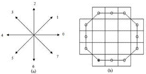
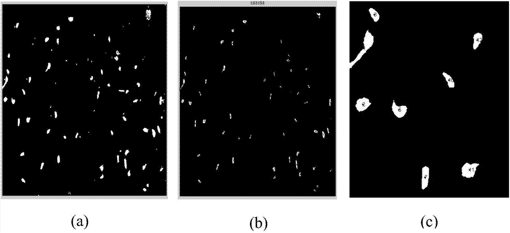
The invention discloses an automatic cutting and counting method for fluorescent microscopic images of retinal cells. The method comprises the following steps: (a) preprocessing, wherein the images are preprocessed and noise points in the images are filtered away; (b) boundary encoding, wherein profiles of the cells are extracted and the extracted profiles are encoded; (c) concave point detection, wherein concave points in the profiles are found and marked; (d) cutting, wherein adherent cells are cut. The method combines a conventional algorithm and various algorithms proposed in modern time, and retains the advantages of simplicity for calculation, low calculation cost and high running efficiency in threshold filtration and edge detection algorithms. At the same time, the method combines Freeman chain code and polygonal concavity and convexity methods, better cuts the adherent cells and enables a cutting result to have relatively good accuracy and relatively high efficiency.

Description

technical field [0001] The design relates to the automatic segmentation and counting method of fluorescent microscopic images of retinal cells, and belongs to the technical field of cell image segmentation and image processing. Background technique [0002] In the process of diagnosis, treatment and repair of diseases, the study of biological cell structure and morphological changes can provide assistance for the diagnosis and treatment of diseases. The most important and difficult thing is the identification and segmentation of cell morphology in biological cell images. Due to the complexity and diversity of cell images, there is currently no fully general segmentation method that can correctly segment all cell images. The segmentation of overlapping and cohesive cells is a major problem in cell image segmentation. In order to improve the accuracy of the analysis results, it is necessary to use a separation algorithm to automatically separate them into individual cells. ...

Claims


Application Information

Patent Timeline
IPC IPC(8): G06K9/00G06K9/36
Inventor 陈新建卢韦华杨磊朱伟芳陈浩宇
Owner SUZHOU UNIV
Why Eureka
Try Eureka