Method for establishing three-dimensional electromagnetic breast simulation model on basis of clinical MRI (magnetic resonance imaging) images

An electromagnetic simulation and breast technology, applied in the field of biomedical detection, can solve problems such as signal attenuation, and achieve the effect of enhancing robustness

Inactive Publication Date: 2015-07-22
TIANJIN UNIV
View PDF2 Cites 10 Cited by
  • Summary
  • Abstract
  • Description
  • Claims
  • Application Information

AI Technical Summary

Problems solved by technology

These tissues will cause a large scattering of the signal, and the gland will cause a large attenuation of the signal, which is undoubtedly a greater challenge for signal processing and imaging

Method used


Image

  • Method for establishing three-dimensional electromagnetic breast simulation model on basis of clinical MRI (magnetic resonance imaging) images
  • Method for establishing three-dimensional electromagnetic breast simulation model on basis of clinical MRI (magnetic resonance imaging) images
  • Method for establishing three-dimensional electromagnetic breast simulation model on basis of clinical MRI (magnetic resonance imaging) images

Examples


Embodiment Construction

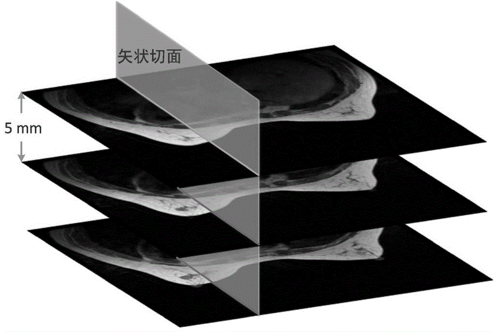
[0025] The present invention first stretches the MRI source images of the whole group of medical images, so that each pixel matches the FDTD grid. Then, all the MRI source images are stacked and interpolated in the direction perpendicular to the image, so that each pixel in the vertical direction is aligned with the FDTD grid. Second, perform boundary recognition on each interpolated cross-section to find the boundary between the interior of the breast and the air. Thirdly, the internal tissue of the breast is discretely divided, and the breast is divided into different tissue categories according to the gray value, and the skin layer is added along the breast boundary. Finally, the numbers and electrical characteristics of different tissues are assigned, so that the model can be applied in the FDTD electromagnetic simulation model. Specific steps are as follows:

[0026] 1. Source image stretching

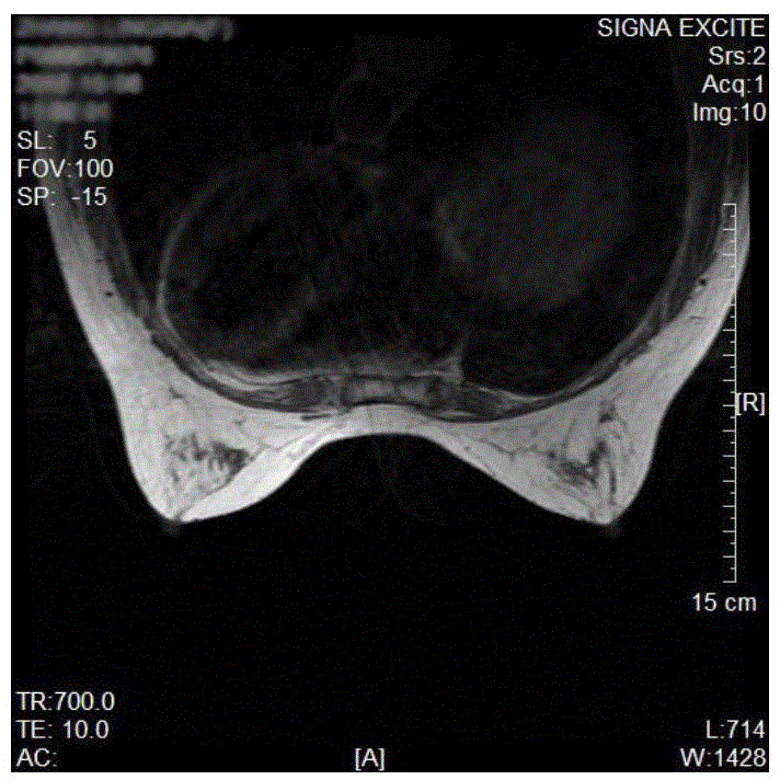
[0027] The T1 source images provided by the hospital basically have a reso...


PUM


Abstract

The invention relates to a method for establishing a three-dimensional electromagnetic breast simulation model on the basis of clinical MRI (magnetic resonance imaging) images. The method includes the step of (1) performing stretching processing on each breast section image by a bicubic interpolation method; (2) stacking breast sections subjected to the stretching processing sequentially to obtain a three-dimensional pixel-point breast array; (3) obtaining a peripheral outline of a breast model; (4) subjecting breast tissue to discrete division; (5) obtaining a peripheral outline of a thoracic cavity, and determining a part, positioned within the peripheral outline of the thoracic cavity, of the breast model to be the thoracic cavity; (6) assigning different electrical property values for different breast tissue obtained in the step (4), and establishing the electromagnetic breast simulation model. The method for establishing the three-dimensional electromagnetic breast simulation model on the basis of the clinical MRI images has the advantages that the established breast model is capable of morphologically reflecting breasts in aspects of shape and internal tissue, such as fat, glands and the thoracic cavity.

Description

Technical field [0001] The invention belongs to the field of biomedical detection and relates to a breast electromagnetic simulation model Background technique [0002] Breast cancer is the malignant tumor disease with the highest incidence rate in women, and its mortality rate ranks first in the mortality rate of malignant tumors in women. The diagnosis of early breast tumors is undoubtedly of decisive significance for improving the treatment rate of breast disease and the long-term survival rate of patients. Currently commonly used detection methods for early breast cancer include mammography, ultrasound imaging technology, computed tomography, nuclear magnetic resonance imaging technology, thermal imaging detection, etc., but many methods have certain shortcomings, such as radiation damage to the human body, The imaging contrast is low and the cost is high. The principle of ultra-broadband electromagnetic wave detection of breast cancer is that different biological tiss...

Claims


Application Information

Patent Timeline
Patent Type & Authority Applications(China)
IPC IPC(8): G06T17/00
Inventor 肖夏王梁宋航
Owner TIANJIN UNIV
Why Eureka
Try Eureka