Fungal keratitis image recognition method based on RX anomaly detection and texture analysis

A fungal keratitis and anomaly detection technology, which is applied in the field of image processing, can solve the problems of decreased diagnostic accuracy, poor performance, and inability to remove background interference very effectively, so as to remove the interference of background information and improve the recognition rate Effect

Active Publication Date: 2015-08-19
SHANDONG UNIV
View PDF4 Cites 4 Cited by
  • Summary
  • Abstract
  • Description
  • Claims
  • Application Information

AI Technical Summary

Problems solved by technology

A large number of medical images have caused two problems: On the one hand, reading the examination images of patients has become a very heavy task for doctors. Due to long-term interpretation of images, doctors are prone to fatigue and distraction, resulting in a decline in the accuracy of diagnosis; On the other hand, it is difficult to guarantee that there will be no missed diagnosis and misdiagnosis by relying on the doctor's own experience, and it is difficult to conduct a consistent quantitative analysis of the imaging data, and the quantitative analysis of medical images is an inevitable requirement for the development of medical imaging
Traditional image preprocessing methods such as brightness transformation, spatial filtering, and histogram processing do not perform very well, and cannot effectively remove background interference.

Method used


Image

  • Fungal keratitis image recognition method based on RX anomaly detection and texture analysis
  • Fungal keratitis image recognition method based on RX anomaly detection and texture analysis
  • Fungal keratitis image recognition method based on RX anomaly detection and texture analysis

Examples


Embodiment Construction

[0058] The present invention will be further described below in conjunction with the accompanying drawings and embodiments.

[0059] Such as Figure 4 As shown, the fungal keratitis image recognition method based on RX anomaly detection and texture analysis includes:

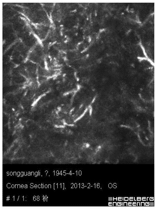
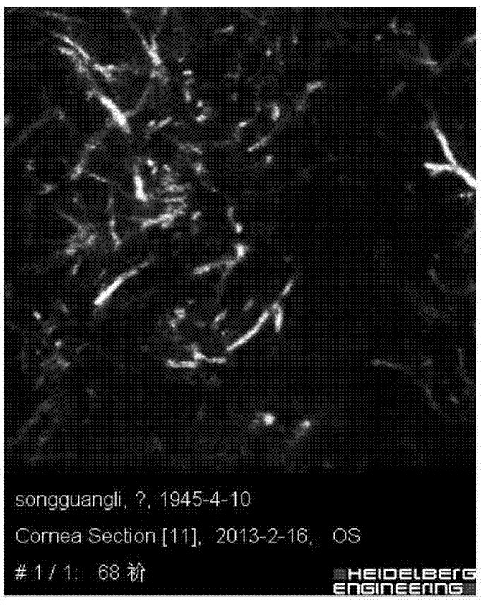
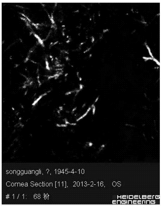
[0060] Step (1): Obtain normal corneal nerve images and hyphae images containing only hyphae as training samples; obtain fundus images of patients with fungal keratitis as test samples;

[0061] Step (2): said step (2) includes step (2-1), step (2-2) and step (2-3) carried out concurrently;

[0062] Step (2-1): performing preprocessing, feature extraction and feature fusion on the normal corneal nerve image in the training sample to obtain the neural feature after the training sample fusion;

[0063] Step (2-2): Preprocessing, feature extraction and feature fusion are performed on the mycelium image that only contains hyphae in the training sample to obtain the hyphae feature after the training sample fusion; ...


PUM


Abstract

The present invention discloses a fungal keratitis image recognition method based on RX anomaly detection and texture analysis. The method comprises a step of obtaining a normal corneal nerve image and a mycelium image which comprises mycelium only as a training sample, a step of obtaining the fundus image of a fungal keratitis patient as a test sample, a step of carrying out preprocessing, feature extraction and feature integration on the normal corneal nerve image in the training sample to obtain a nerve feature after training sample integration, a step of carrying out preprocessing, feature extraction and feature integration on the mycelium image which comprises the mycelium only in the training sample to obtain a mycelium feature after the training sample integration, a step of carrying out preprocessing, feature extraction and feature integration on the image in the test sample to obtain the nerve feature after test sample integration and the mycelium feature after the test sample integration, and a step of recognizing the nerve and mycelium in the test sample.

Description

technical field [0001] The invention belongs to the technical field of image processing, in particular to an image recognition method for fungal keratitis based on RX abnormality detection and texture analysis. Background technique [0002] Fungal keratitis is an infectious corneal disease with a high blinding rate caused by pathogenic fungi. Due to its rapid development, it is very easy to cause serious consequences such as corneal perforation, hypopyon, endophthalmitis, etc., so it is extremely harmful , the early diagnosis and treatment of this disease is particularly important. Confocal microscope is a new type of non-invasive corneal imaging examination instrument, which can scan and image the cornea from the four-dimensional (three-dimensional space and time) level in vivo, and provide high-definition and magnification images of all layers of the cornea , allowing people to directly observe and study the pathophysiology of living corneas at the cellular level. [000...

Claims


Application Information

Patent Timeline
Patent Type & Authority Applications(China)
IPC IPC(8): G06K9/62
CPCG06F18/24133G06F18/253G06F18/214
Inventor 刘治刘红彤张明高张海霞吴雪莲陶远
Owner SHANDONG UNIV
Why Eureka
Try Eureka