In-vitro separation and cultivation method of deer dermal papilla cells and application thereof

A technique for separating and culturing dermal papilla cells, which is applied to artificial cell constructs, animal cells, vertebrate cells, etc., can solve the problems of difficulty in collecting dermal papillae, affecting the collection of dermal papillae, and uneven digestion of dermal papillae, etc. Separation efficiency, improving the success rate of culture, and improving the effect of adherence rate

Inactive Publication Date: 2015-09-16
INST OF SPECIAL ANIMAL & PLANT SCI OF CAAS
View PDF2 Cites 3 Cited by
  • Summary
  • Abstract
  • Description
  • Claims
  • Application Information

AI Technical Summary

Problems solved by technology

Enzyme digestion directly digests the skin tissue containing hair follicles, resulting in a lot of skin tissue fragments, making it difficult to collect dermal papilla, resulting in uneven digestion of dermal papilla, affecting the collection of dermal papilla

Method used


Image

  • In-vitro separation and cultivation method of deer dermal papilla cells and application thereof
  • In-vitro separation and cultivation method of deer dermal papilla cells and application thereof
  • In-vitro separation and cultivation method of deer dermal papilla cells and application thereof

Examples


Embodiment 1

[0026] Embodiment 1: Utilize the method for human scalp dermal papilla cells to isolate and cultivate deer fur dermal papilla cells

[0027] 1. The skin of the live deer is collected and treated aseptically.

[0028] 2. Cut at the dermis-subcutaneous tissue junction. Microscopic tweezers lightly press the subcutaneous fat containing the middle and lower part of the hair follicle to partially extrude the tip of the cut edge of the hair follicle, and then use another pair of micro tweezers to hold the tip of the cut edge to completely pull out the middle and lower part of the hair follicle from the subcutaneous fat. The extracted hair follicles were placed in a petri dish filled with sterile D-hanks solution.

[0029] 3. Under the microscope, use micro-tweezers to clamp the hair follicle near the hair bulb. Since the epidermis and dermis are closely connected, loose hair matrix cells and melanocytes can only migrate to the hair bulb. The hair bulb forms a spherical shape with ...

Embodiment 2

[0033] Specific steps are as follows:

[0034] 1. Cut the skin of the deer collected from the live body into 1-2 strips of 0.5x3cm, bring them to the laboratory, wash off the blood on the tissue pieces with PBS, and carry out disinfection treatment.

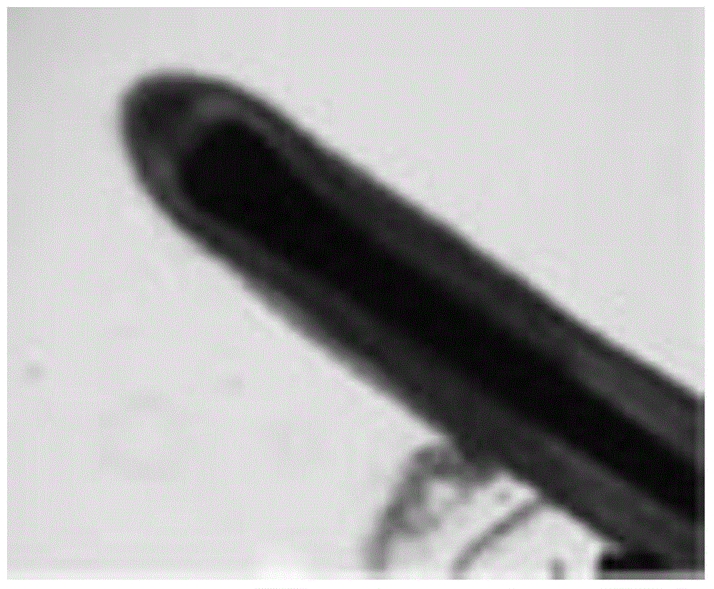
[0035] 2. Cut the leather strip into 0.7mm slices along the growth direction of the hair follicles, and pull out the complete hair follicles from each slice under a stereomicroscope, and collect them in a petri dish ( figure 1 ).

[0036] 3. Digest the hair follicles with 100U / ml type II collagenase at 37°C for 30min. Discard the digestion solution and add DMEM medium.

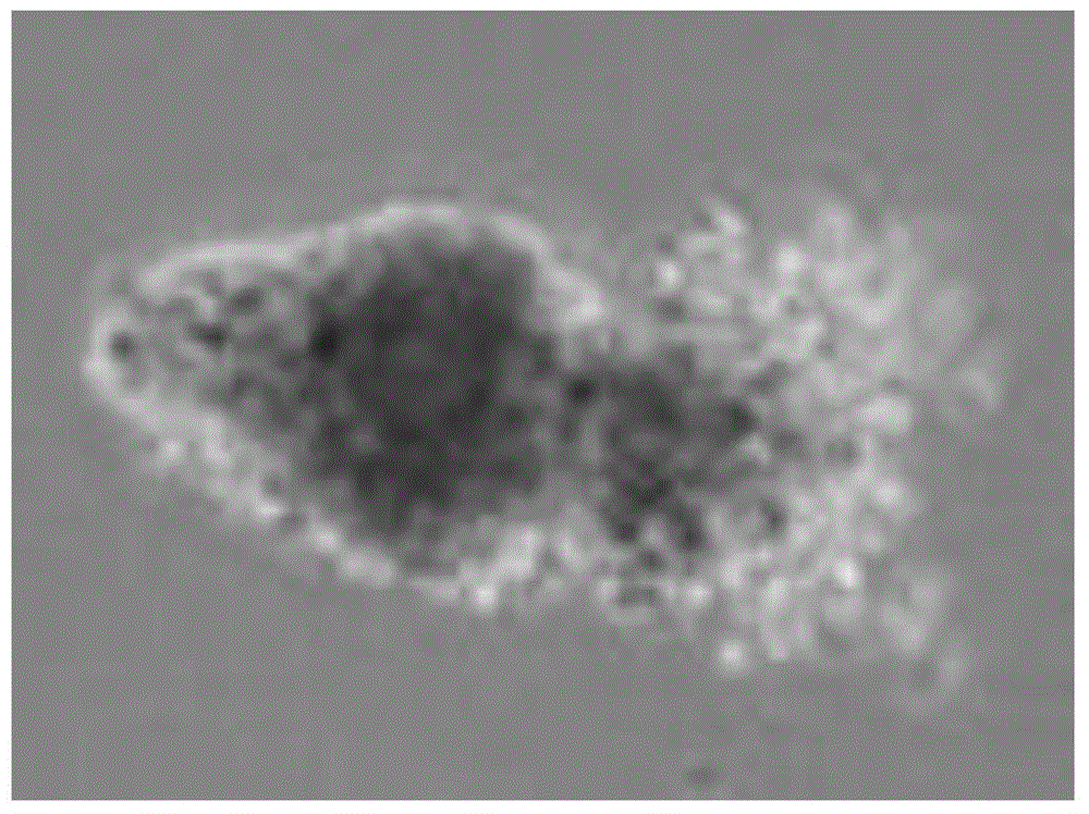
[0037] 4. Under a stereomicroscope, use the wedge-shaped surface of the needle tip of a 1mL syringe to cut off the bulb of the hair follicle containing the hair papilla, use microscopic forceps to open the root sheath of the hair to expose the hair papilla, and use the needle tip of the syringe to cut the hair papilla and the hair matrix and / or the connectio...


PUM


Abstract

The invention discloses an in-vitro separation and cultivation method of deer dermal papilla cells and application thereof, and belongs to the technical field of cytobiology. The in-vitro separation and cultivation method comprises the steps that the complete hair follicle in deer skin is separated under an integrated microscope; then collagenase is used for digesting the hair follicle; a hair bulb containing a papilla is cut off; a hair root sheath is stripped to obtain the papilla; the papilla is collected; then the collagenase is used for digesting a basilar membrane; and the papilla cells are acquired after development. The in-vitro separation and cultivation method has the advantages that the cultivation success rate is very high, and the adherence rate of the papilla and the cell yield are obviously improved. By the adoption of the cultivation method, about 100 pure dermal papillae can be obtained within about four hours, and a larger number of dermal papilla cells can be obtained when the dermal papillae are subjected to inoculated culture for about seven days. The in-vitro separation and cultivation method is suitable for in-vitro separation and cultivation of the deer dermal papilla cells.

Description

technical field [0001] The invention relates to an in vitro separation and cultivation method and application of deer dermis dermal papilla cells, belonging to the technical field of cell biology. Background technique [0002] At present, the dermal papilla cell culture technology is mainly aimed at the dermal papilla cells of the human scalp and the dermal papilla cells of the mouse beard. There is no culture technology for the dermal papilla cells of the deer skin. Compared with the human hair and the beard of the mouse, the deer hair is thinner, The dermal dermal papilla of deer is small, and there are certain differences between deer skin structure and human scalp. Therefore, the existing method for culturing dermal dermal papilla cells is not suitable for deer skin. The existing methods for culturing dermal papilla cells mainly include microdissection, enzymatic digestion, enzymatic digestion and filtration, etc. The adherence rate of dermal papilla isolated by microdi...

Claims


Application Information

Patent Timeline
Patent Type & Authority Applications(China)
IPC IPC(8): C12N5/071
Inventor 孙红梅李春义褚文辉路晓赵海平
Owner INST OF SPECIAL ANIMAL & PLANT SCI OF CAAS
Why Eureka
Try Eureka