

Fundus OCT imaging method and system for binocular stereo vision three-dimensional imaging
A technology of binocular stereo vision and imaging system, applied in the field of blood flow imaging
- Summary
- Abstract
- Description
- Claims
- Application Information
AI Technical Summary
Problems solved by technology
Method used
Image

Examples
Embodiment Construction
[0079] Embodiments of the present invention will be described in detail below with reference to the accompanying drawings. When describing the present invention, if the description of the notification function or structure related to the invention is unnecessary, the description of this part can be omitted. Also, the functions described below are all functions defined in consideration of the present invention, and the functions are variable according to the user's intention or practice, so the definition should be determined based on the entire content of this specification.
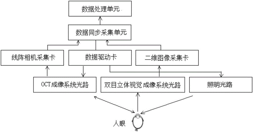
[0080] The block diagram of the OCT imaging system is as follows: figure 1 As shown, by the optical path (OCT imaging system optical path, binocular stereo vision system imaging optical path and lighting optical path), computer internal boards (data drive card, line array camera acquisition card and two-dimensional image acquisition card) and algorithm (data synchronous acquisition unit and data process...
PUM
 Abstract
Description
Claims
Application Information