Blood vessel extraction method

An extraction method and blood vessel technology, applied in the field of medical image processing, can solve the problems of over-segmentation, inability to extract small branches of blood vessels, fast calculation speed, etc., and achieve the effect of complete extraction

Inactive Publication Date: 2015-10-14
SHANGHAI UNITED IMAGING HEALTHCARE
View PDF2 Cites 28 Cited by
  • Summary
  • Abstract
  • Description
  • Claims
  • Application Information

AI Technical Summary

Problems solved by technology

The calculation speed of this method is fast, because it is related to the threshold value, the robustness is poor
When the threshold range is too small, it is impossible to extract small branches of blood vessels. When the threshold range is too large, some tissues such as bones will be regarded as blood vessels, resulting in over-segmentation

Method used


Image

  • Blood vessel extraction method
  • Blood vessel extraction method
  • Blood vessel extraction method

Examples


Embodiment Construction

[0026] In the following description, numerous specific details are set forth in order to provide a thorough understanding of the present invention. However, the present invention can be implemented in many other ways different from those described here, and those skilled in the art can make similar extensions without violating the connotation of the present invention, so the present invention is not limited by the specific implementations disclosed below.

[0027] Secondly, the present invention is described in detail by means of schematic diagrams. When describing the embodiments of the present invention in detail, for convenience of explanation, the schematic diagrams are only examples, which should not limit the protection scope of the present invention.

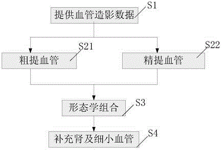
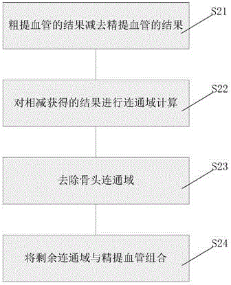
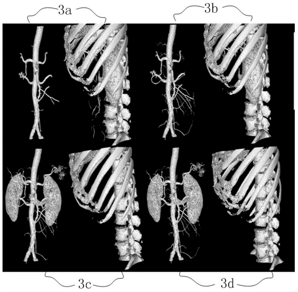
[0028] The present invention provides a method for extracting blood vessels, which is characterized in that it includes: reading angiographic data, and using the region growing method to obtain the first result for the dat...


PUM


Abstract

The invention provides a blood vessel extraction method. The method comprises steps of reading angiography data; adopting a region growing method on the data to obtain a first result; adopting a level set method to obtain a second result; performing morphological combination according to the first result and the second result; and performing blood vessel extraction of the angiography data. According to the invention, advantages of the region growing method based on the threshold and the gradient and the level set method based on the radius and the gradient are effectively combined, and region growing of bones and blood vessels are flexibly utilized to assist the blood vessel extraction, so that the complete extraction of blood vessels can be achieved.

Description

technical field [0001] The invention relates to the field of medical image processing, in particular to a blood vessel extraction method. Background technique [0002] Angiography (CTA) is one of the important methods for diagnosing vascular diseases, and it is mainly used in clinical diagnosis and treatment of various vascular diseases, such as aneurysm, vascular stenosis, and vascular calcification. Segmenting blood vessels from other tissues is a critical step in angiographic techniques. Due to the overlap of CT values ​​of blood vessels and other tissues such as bones in CTA images, the current main blood vessel segmentation methods such as region growth and dynamic contours are easy to mistake bones and other tissues for blood vessel extraction, resulting in over-segmentation or small blood vessels as non-vessels Tissue removal, resulting in incomplete segmentation. [0003] Document 1: P.T.Vieco, W.P.Shuman, G.F.Alsofrom and C.E.Gross.Detection of circle of Willis an...

Claims


Application Information

Patent Timeline
Patent Type & Authority Applications(China)
IPC IPC(8): G06T7/00
Inventor 毛玉妃马杰延李程
Owner SHANGHAI UNITED IMAGING HEALTHCARE
Why Eureka
Try Eureka