Image processing method for detecting pulmonary tuberculosis focus in chest X-ray DR film

An image processing and pulmonary tuberculosis technology, applied in the field of image processing, can solve the problems of large screening workload, difficult time for doctors, large data volume, etc., and achieve the effect of easy acceptance, reduce workload, and improve recognition rate.

Inactive Publication Date: 2015-12-09
SICHUAN UNIV
View PDF3 Cites 16 Cited by
  • Summary
  • Abstract
  • Description
  • Claims
  • Application Information

AI Technical Summary

Problems solved by technology

Doctors in grass-roots medical and health institutions must supervise and visit regularly; large-scale resident physical examinations generate too much data, and the workload...

Method used


Image

  • Image processing method for detecting pulmonary tuberculosis focus in chest X-ray DR film

Examples


Embodiment Construction

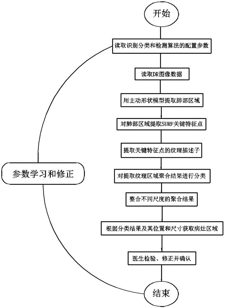
[0036] The present invention will be further described below in conjunction with the accompanying drawings and specific embodiments.

[0037] An image processing method for detecting pulmonary tuberculosis lesions in a chest X-ray DR film, comprising the following steps:

[0038] Extract images of lung regions through active shape models;

[0039] Extract the key feature points of SURF in the lung area image;

[0040] Extract texture descriptors of key feature points;

[0041] Specify the category of key feature points, use the k-means clustering algorithm to aggregate the texture description sub-clusters, and classify and mark the aggregation results;

[0042] Set a set of size values, and aggregate the texture annotations of the lung region images in order of size values ​​from small to large;

[0043] Delete the categories whose number of samples is less than 5% of the total in the clustering results, so as to keep only the main categories in the clustering results, remove...


PUM


Abstract

The invention provides an image processing method for detecting a pulmonary tuberculosis focus in a chest X-ray DR film. The image processing method comprises the following steps of extracting a lung area image through an active shape model; extracting SURF key feature points of a lung area image; extracting texture descriptors of the key feature points; assigning the kind of the key feature points; performing clustering and combination on the texture descriptors according to a k-means clustering algorithm, and performing classified making on a combination result; setting a group of dimension values, and combining the texture marks of the lung area image according to a sequence from a least dimension value to a minimum dimension value; and deleting the class of which the number of samples is smaller than 5% of the total number in the combining result, thereby only reserving main classes in the combination result, eliminating abnormities and obtaining an image processing result. The image processing method can be used for automatically processing the DR films. The image processing method is applied for screening the pulmonary tuberculosis. The lung DR films which may represent the pulmonary tuberculosis can be screened. The image processing method can be used for large-number pulmonary tuberculosis screening. The image processing method has advantages of high screening efficiency and objective and stable conclusion.

Description

technical field [0001] The invention relates to an image processing method, in particular to an image processing method for detecting pulmonary tuberculosis lesions in chest X-ray DR films. Background technique [0002] Tuberculosis is caused by Mycobacterium tuberculosis, which is easily transmitted by droplets or even aerosols in the air; an infectious tuberculosis patient can infect 10 to 15 people on average in a year; BCG vaccination of newborns cannot prevent adult diseases, tuberculosis The main body of patients is young and middle-aged people, once the disease occurs, it will lead to the loss of family and social labor force; the World Health Organization pointed out that tuberculosis is an important public health problem in the world. Globally, tuberculosis kills 1.4 million people every year; in China, there are currently 5 million active pulmonary tuberculosis 50,000 people die from tuberculosis every year, which is equivalent to 1 death every 10 minutes; accordin...

Claims


Application Information

Patent Timeline
IPC IPC(8): G06T7/00G06K9/46G06K9/62
CPCG06T7/0012G06T2207/30061G06T2207/10116G06V10/462G06F18/23213
Inventor 王俊峰唐鹏姬郁林李虹
Owner SICHUAN UNIV
Why Eureka
Try Eureka