Biological protein two-dimensional nano film prepared by lysozyme and preparation method thereof

A two-dimensional nanotechnology, protein technology, applied in nanotechnology and other directions, can solve the problems of difficult realization of macroscopic structure and complex synthesis route, and achieve the effect of small surface roughness, high transparency and simple preparation method

Active Publication Date: 2018-01-12
SHAANXI NORMAL UNIV
View PDF2 Cites 2 Cited by
  • Summary
  • Abstract
  • Description
  • Claims
  • Application Information

AI Technical Summary

Problems solved by technology

It is more difficult to achieve large-scale (more than centimeter-scale) macroscopic tissues formed by self-assembly of microscopic building blocks, and complex synthetic routes are often involved to obtain target products or peptides

Method used


Image

  • Biological protein two-dimensional nano film prepared by lysozyme and preparation method thereof
  • Biological protein two-dimensional nano film prepared by lysozyme and preparation method thereof
  • Biological protein two-dimensional nano film prepared by lysozyme and preparation method thereof

Examples


Embodiment 1

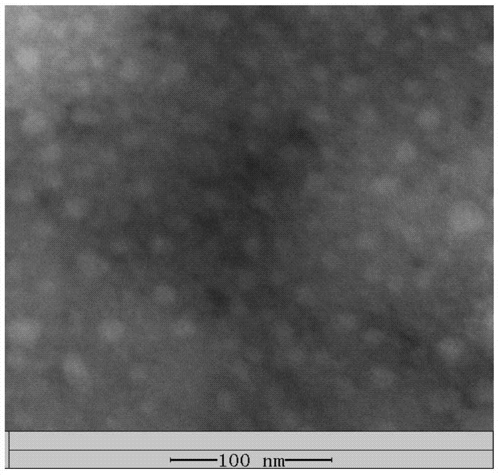
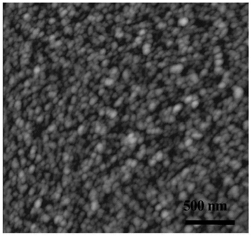
[0025] Add 0.1433g of tris(2-carboxyethyl)phosphine into 10mL of 10mmol / L tris(hydroxymethylaminomethane) buffer solution with a pH value of 7.4, adjust the pH value to 5.0 with NaOH, and prepare 50mmol / L of tris(2- Carboxyethyl) phosphine tris buffer solution; 20mg lysozyme was added to 10mL 10mmol / L tris buffer solution with a pH value of 7.4 to prepare 2mg / mL trihydroxymethyl aminomethane buffer solution of lysozyme Methylaminomethane buffer solution; the tris (2-carboxyethyl)phosphine tris buffer solution of 10mL 50mmol / L is mixed with the tris buffer solution of lysozyme of 10mL2mg / mL, Stand at room temperature for 50 minutes to form a thin film on the surface of the mixture (such as figure 1 Shown), that is, biological protein two-dimensional nano film, its thickness is about 50nm. Depend on figure 2 and image 3 It can be seen that the two-dimensional nano-film of biological protein is formed by the self-assembly of nanoparticles with a particle size of about 30nm-5...

Embodiment 2

[0027] Add 0.1147g of tris(2-carboxyethyl)phosphine into 10mL of 10mmol / L tris(hydroxymethylaminomethane) buffer solution with a pH value of 7.4, adjust the pH value to 5.0 with NaOH, and prepare 40mmol / L of tris(2- Carboxyethyl) phosphine tris buffer solution; 50mg lysozyme was added into 10mL 10mmol / L tris buffer solution with a pH value of 7.4 to prepare 5 mg / mL of lysozyme tris Methylaminomethane buffer solution; the tris (2-carboxyethyl) phosphine tris buffer solution of 10mL 40mmol / L is mixed with the tris buffer solution of lysozyme of 10mL5mg / mL, After standing still at room temperature for 50 minutes, a thin film, that is, a two-dimensional nano-film of biological protein, is formed on the surface of the mixed liquid, and its thickness is about 60 nm.

Embodiment 3

[0029] Add 0.1433g of tris(2-carboxyethyl)phosphine into 10mL of 10mmol / L tris(hydroxymethylaminomethane) buffer solution with a pH value of 7.4, adjust the pH value to 6.0 with NaOH, and prepare 50mmol / L of tris(2- Carboxyethyl) phosphine tris buffer solution; 20mg lysozyme was added to 10mL 10mmol / L tris buffer solution with a pH value of 7.4 to prepare 2mg / mL trihydroxymethyl aminomethane buffer solution of lysozyme Methylaminomethane buffer solution; the tris (2-carboxyethyl)phosphine tris buffer solution of 10mL 50mmol / L is mixed with the tris buffer solution of lysozyme of 10mL2mg / mL, After standing still at room temperature for 50 minutes, a thin film, that is, a two-dimensional nano-film of biological protein, is formed on the surface of the mixed liquid, and its thickness is about 60 nm.


PUM

PropertyMeasurementUnit
thicknessaaaaaaaaaa
thicknessaaaaaaaaaa
particle diameteraaaaaaaaaa

Abstract

The invention discloses a method for preparing a two-dimensional nano-film of biological protein by using lysozyme. The method utilizes the phase transition reaction of lysozyme molecules and tris(2-carboxyethyl)phosphine hydrochloride to produce nanoparticles at the gas-liquid interface. A layer of biological protein two-dimensional nano film is formed by surface interface induced self-assembly, or the substrate is directly contacted with the surface of the solution during the preparation of the film, so that the nanoparticles generated by the phase transition of lysozyme are directly on the liquid-solid surface through surface interface induction Self-assembly forms a two-dimensional nano-film, that is, grows in situ on the surface of the substrate. The preparation method of the present invention is simple and environment-friendly, the thickness of the prepared biological protein two-dimensional nano-film is about 30-80nm, the surface roughness is small, the transparency is high, and it has good adhesion, and can be used for substrate surface Make modifications.

Description

technical field [0001] The invention belongs to the technical field of preparation of biological protein two-dimensional nano film, and in particular relates to a biological protein two-dimensional nano film prepared by using lysozyme and a preparation method thereof. Background technique [0002] Among the various physiological functions in nature, the self-assembly of protein aggregates is particularly common. For example: amelogenin can self-assemble to form nanospheres, and these nanosphere systems assemble to form a ribbon structure. This self-assembled structure plays a key template role by controlling the growth of hydroxyapatite crystals to achieve Biomineralization of tooth enamel. Nowadays, protein two-dimensional nanofilms formed by self-assembly have attracted extensive attention from the scientific community. Especially during the phase transition, supramolecular aggregates are formed by the spontaneous aggregation of hydrophobic particles, thereby forming a t...

Claims


Application Information

Patent Timeline
Patent Type & Authority Patents(China)
IPC IPC(8): C08J5/18C08L89/00B82Y40/00
Inventor 杨鹏哈媛王德辉
Owner SHAANXI NORMAL UNIV
Why Eureka
Try Eureka