Medical image processing device, method for operating the same, and endoscope system
一种处理装置、医用图像的技术,应用在内窥镜系统领域,能够解决难以强调萎缩部色差等问题
- Summary
- Abstract
- Description
- Claims
- Application Information
AI Technical Summary
Problems solved by technology
Method used
Image

Examples
no. 1 approach
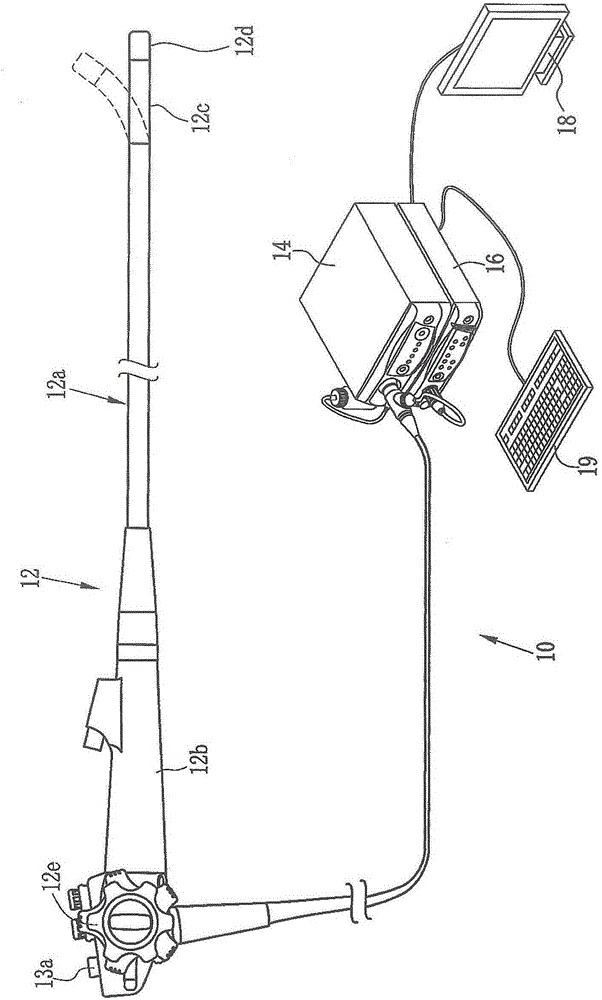
[0070] Such as figure 1 As shown, the endoscope system 10 of the first embodiment includes an endoscope 12 , a light source device 14 , a processor device 16 , a monitor 18 (display unit), and a console 19 . The endoscope 12 is optically connected to a light source device 14 and electrically connected to a processor device 16 . The endoscope 12 has an insertion portion 12a inserted into the subject, an operation portion 12b provided at a base end portion of the insertion portion 12a, and a curved portion 12c and a distal end portion 12d provided on the distal side of the insertion portion 12a. The bending part 12c is bent by operating the angle knob 12e of the operating part 12b. Accompanied by this bending movement, the front end portion 12d faces a desired direction.
[0071] Moreover, the operation part 12b is provided with the mode switch SW13a other than the angle knob 12e. The mode switch SW13a is used for switching operations among four modes of the normal observatio...
no. 2 approach
[0149] In the second embodiment, instead of the LEDs 20 a to 20 d of four colors shown in the first embodiment, a laser light source and a phosphor are used to illuminate an observation object. Other than that, it is the same as the first embodiment.
[0150] Such as Figure 40 As shown, in the endoscope system 100 of the second embodiment, instead of the LEDs 20a to 20d of four colors, a blue laser light source (in the case of Figure 40 marked as "445LD") 104, and a blue-violet laser light source that emits a blue-violet laser with a center wavelength of 405±10nm (in Figure 40 marked as "405LD")106. The light emitted from the semiconductor light emitting elements of these light sources 104 and 106 is separately controlled by the light source control unit 108 so that the light quantity ratio of the blue laser light source 104 and the blue laser light source 106 can be freely changed.
[0151] In the normal observation mode, the light source control unit 108 drives the blu...
no. 3 approach
[0157] In the third embodiment, instead of the four-color LEDs 20 a to 20 d shown in the first embodiment, a broadband light source such as a xenon lamp and a rotary filter are used to illuminate an observation object. In addition, instead of the color imaging sensor 48, a monochrome imaging sensor is used to image the observation object. Other than that, it is the same as the first embodiment.
[0158] Such as Figure 43 As shown, in the endoscope system 200 of the third embodiment, in the light source device 14, a broadband light source 202, a rotary filter 204, and a filter switching unit 205 are provided instead of the LEDs 20a to 20d of four colors. . In addition, instead of the color imaging sensor 48, the imaging optical system 30b is provided with a monochrome imaging sensor 206 not provided with a color filter.
[0159] The broadband light source 202 is a xenon lamp, a white LED, etc., and emits white light with a wavelength range from blue to red. The rotary filt...
PUM
 Abstract
Description
Claims
Application Information
 - Generate Ideas
- Intellectual Property
- Life Sciences
- Materials
- Tech Scout
- Unparalleled Data Quality
- Higher Quality Content
- 60% Fewer Hallucinations
Browse by: Latest US Patents, China's latest patents, Technical Efficacy Thesaurus, Application Domain, Technology Topic, Popular Technical Reports.
© 2025 PatSnap. All rights reserved.Legal|Privacy policy|Modern Slavery Act Transparency Statement|Sitemap|About US| Contact US: help@patsnap.com