Method for automatically identifying and distinguishing eye fundus images

A fundus image, automatic recognition technology, applied in image analysis, image enhancement, image data processing and other directions, can solve the problems of difficult reading, huge reading volume, hindering the development of disciplines, etc., to improve the reading efficiency and reduce the probability of errors. Effect

Active Publication Date: 2016-01-13
成都银海启明医院管理有限公司
View PDF4 Cites 27 Cited by
  • Summary
  • Abstract
  • Description
  • Claims
  • Application Information

AI Technical Summary

Problems solved by technology

[0002] Fundus retinal images can be fully and clearly obtained by fundus photography equipment. Due to the different angles of the camera when taking pictures, or because of the needs of lesion locations, fundus images of different fundus regions are often obtained. The existing partitioning methods are usually divided into nasal and retinal regions centered on the optic disc. In the upper, lower nasal, superior temporal and inferior temporal areas, researchers or doctors study the fundus images, and diagnose and grade related diseases according to the characteristics and severity of lesions in each fundus area. When studying, it is necessary to first determine the area to whi...

Method used


Image

  • Method for automatically identifying and distinguishing eye fundus images
  • Method for automatically identifying and distinguishing eye fundus images
  • Method for automatically identifying and distinguishing eye fundus images

Examples


Embodiment Construction

[0042] The present invention is specifically described below by the examples, the examples are only used to further illustrate the present invention, can not be interpreted as the limitation of the protection scope of the present invention, some non-essential improvements made by those skilled in the art according to the content of the present invention And adjustments also belong to the protection scope of the present invention.

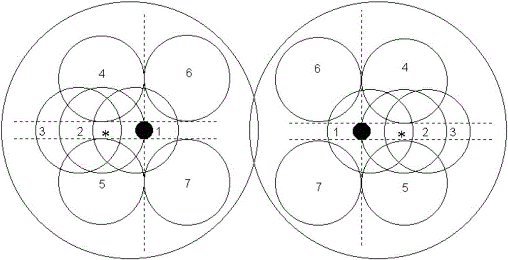
[0043] The classification definition of the seven areas of the fundus image:

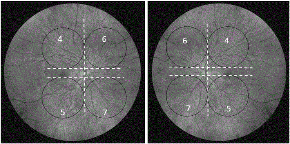
[0044] like figure 1 and figure 2 as shown, figure 1 It is a schematic diagram of the seven zones of the fundus of the present invention; the figure includes a schematic diagram of the zones of the left and right fundus, and the positions of zones 1 to 7 are indicated by circles in the figure. The black dot near the center is the optic disc, and the asterisk near the center* It is the macula, and each number in the figure indicates the number of each area. figure 2 ...


PUM


Abstract

The invention discloses a method for automatically identifying and distinguishing eye fundus images. The method comprises the following steps: obtaining black and white or colored eye fundus image pictures by using eye fundus photographic equipment, and storing the black and white or colored eye fundus image pictures according to a left eye or a right eye; dividing the eye fundus image of the left eye or the right eye into seven regions, which are respectively a first region-optic disk region, a second region-macular region, a third region- macular temporal region, a fourth region- area temporalis superior, a fifth region- area temporalis inferior, a sixth region- superior nasal region and a seventh region- inferior nasal region; and automatically judging that the collected eye fundus image belongs to one of the seven eye fundus regions through the computer graphics according to the optic disk, macular and vascular network information in the eye fundus image. The method disclosed by the invention is used for introducing automatic computer image identification into the processing of the images of the seven eye fundus regions, and extracting feature structures of different eye fundus regions to serve as reference for automatically locating and collecting the eye fundus image, and providing a brand new manner for researchers or film reading doctors to research and read the images of the seven eye fundus regions, so as to greatly improve the film reading efficiency and reduce the error probability.

Description

technical field [0001] The invention relates to a fundus image image processing technology based on computer image recognition technology, in particular to a fundus image automatic recognition partition method. Background technique [0002] Fundus retinal images can be fully and clearly obtained by fundus photography equipment. Due to the different angles of the camera when taking pictures, or because of the needs of lesion locations, fundus images of different fundus regions are often obtained. The existing partitioning methods are usually divided into nasal and retinal regions centered on the optic disc. In the upper, lower nasal, superior temporal and inferior temporal areas, researchers or doctors study the fundus images, and diagnose and grade related diseases according to the characteristics and severity of lesions in each fundus area. When studying, it is necessary to first determine the area to which the obtained fundus image belongs. Under the condition that the fun...

Claims


Application Information

Patent Timeline
IPC IPC(8): G06T7/00
CPCG06T7/0012G06T2207/10004G06T2207/10024G06T2207/30041
Inventor 满文钢王竞段俊国王蕾陈洁厉元杰
Owner 成都银海启明医院管理有限公司
Why Eureka
Try Eureka