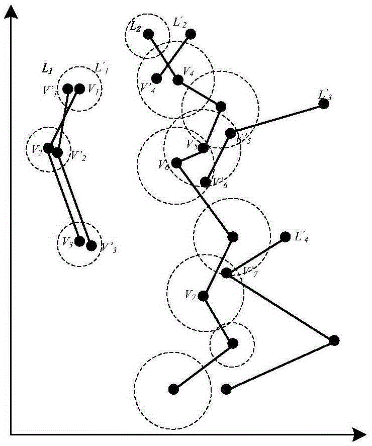
Medical image classification method based on KAP digraph model
A medical image and classification method technology, applied in the field of medical information, can solve the problems of high time complexity and low classification accuracy, and achieve the effects of reducing extraction time, improving representativeness, and improving practical value
- Summary
- Abstract
- Description
- Claims
- Application Information
AI Technical Summary
Problems solved by technology
Method used
Image
Examples
Embodiment 1
[0039] First preprocess the medical image:
[0040] 1. Extract the ROI region from each original brain CT image in the original image library;
[0041] 2. Intercept the ROI area and correct it;
[0042] 3. Calculate the trough distribution of the gray histogram in the ROI region of the image, and obtain the trough table of the gray histogram;
[0043] 4. According to the threshold value set in the valley table, the texture is extracted multiple times from the image to obtain a multi-level texture image;
[0044] 5. Finally, the multi-level texture image is normalized into an image whose size is COLUMN×ROW;
[0045] 6. Extract the corner points of the texture image;
[0046] 7. Store the coordinates of the extracted corner points in the corresponding coordinate queue;
[0047] Save the preprocessed image in the corresponding database. After the above process, each original image corresponds to a texture corner point storage queue. First, use the corner points in the storag...
PUM
 Abstract
Description
Claims
Application Information
 - R&D
- Intellectual Property
- Life Sciences
- Materials
- Tech Scout
- Unparalleled Data Quality
- Higher Quality Content
- 60% Fewer Hallucinations
Browse by: Latest US Patents, China's latest patents, Technical Efficacy Thesaurus, Application Domain, Technology Topic, Popular Technical Reports.
© 2025 PatSnap. All rights reserved.Legal|Privacy policy|Modern Slavery Act Transparency Statement|Sitemap|About US| Contact US: help@patsnap.com


