Tissue elasticity imaging method and graphics processor

A graphics processor, elastography technology, applied in ultrasonic/sonic/infrasound image/data processing, organ motion/change detection, ultrasonic/sonic/infrasonic Permian technology, etc. and other problems to achieve the effect of improving processing speed and ensuring accuracy

Active Publication Date: 2016-02-10
海斯凯尔(山东)医学科技有限公司
View PDF4 Cites 6 Cited by
  • Summary
  • Abstract
  • Description
  • Claims
  • Application Information

AI Technical Summary

Problems solved by technology

[0004] In the process of ultrasonic elastography processing of tissue, it mainly involves the process of estimating the displacement and strain of the tissue under a certain excitation, and the calculation amount is relatively large. Among them, the cross-correlation algorithm in the displacement estimation consumes a lot of time

Method used


Image

  • Tissue elasticity imaging method and graphics processor
  • Tissue elasticity imaging method and graphics processor
  • Tissue elasticity imaging method and graphics processor

Examples


Embodiment Construction

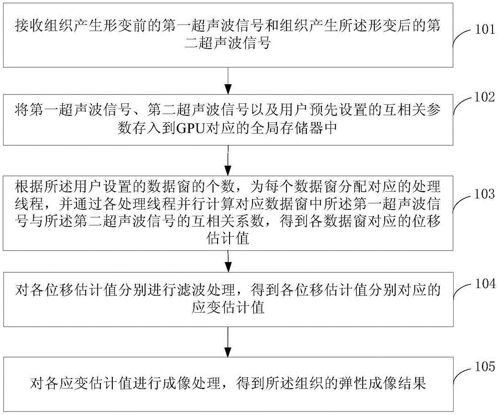
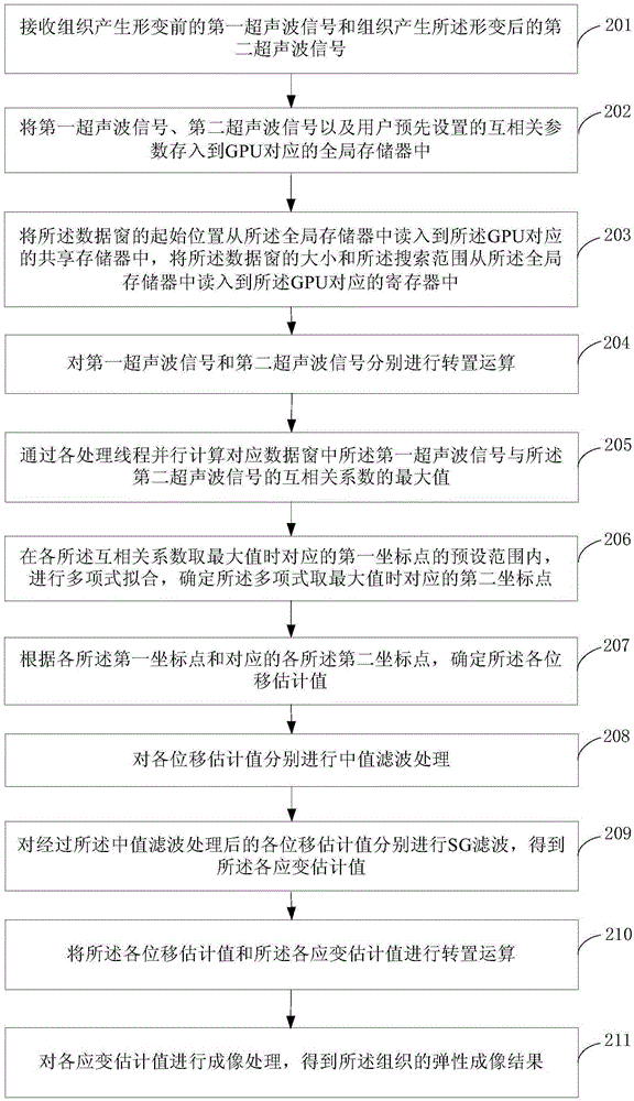
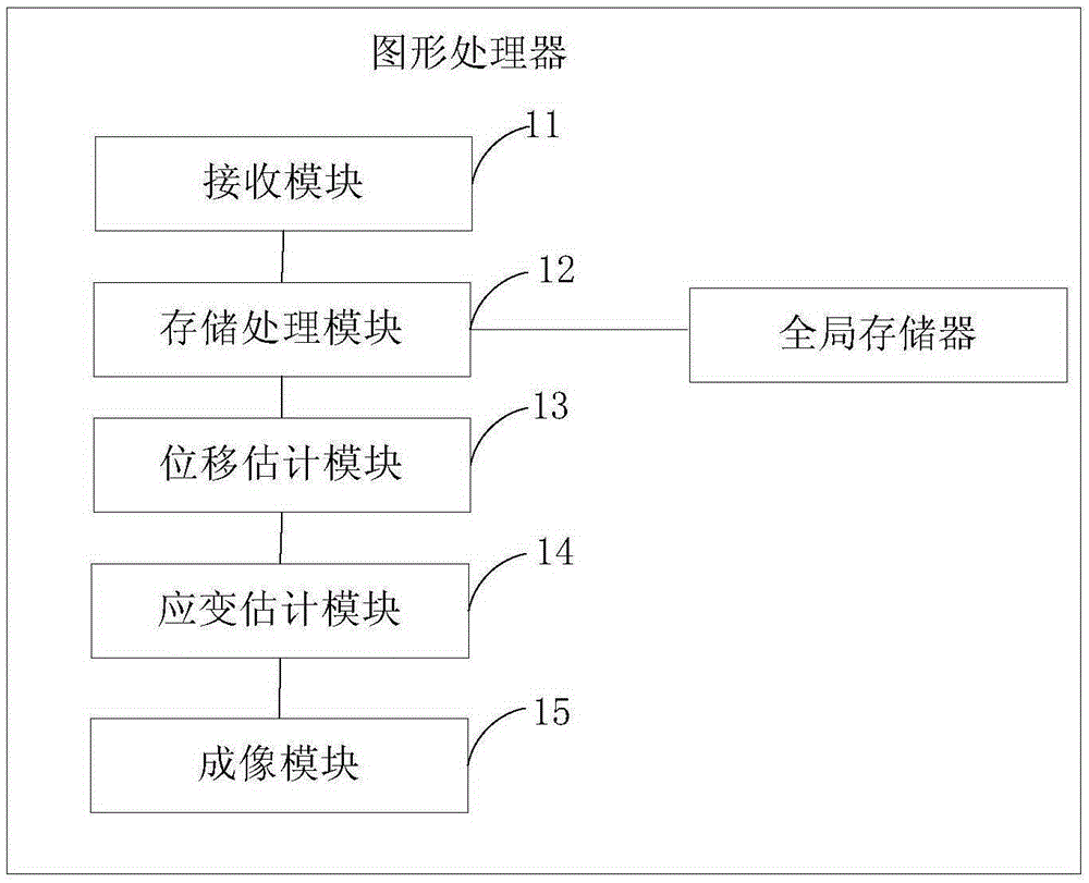
[0023] figure 1 It is a flow chart of Embodiment 1 of the tissue elasticity imaging method of the present invention. In this embodiment, the tissue elasticity imaging method is executed by a Graphic Processing Unit (GPU for short), and the GPU is set in the elasticity detection device, such as figure 1 As shown, the tissue elastography method includes:

[0024] Step 101, receiving a first ultrasonic signal before tissue deformation and a second ultrasonic signal after tissue deformation.

[0025] Step 102, storing the first ultrasonic signal, the second ultrasonic signal and the cross-correlation parameters preset by the user into the global memory corresponding to the GPU.

[0026] In the process of tissue elastography, it is necessary to excite shear waves in the tissue, for example, to excite shear waves in the tissue through mechanical vibration. In order to measure the propagation characteristics of the shear wave in the tissue to obtain the elastic parameters of the ti...


PUM


Abstract

The invention provides a tissue elasticity imaging method and a graphics processor. The tissue elasticity imaging method comprises the following steps: receiving first ultrasonic signals and second ultrasonic signals before and after tissue deformation; storing the first ultrasonic signals, the second ultrasonic signals, parameters contained in a cross-correlation algorithm into a global storage corresponding to a GPU; allocating corresponding processing threads for each data window according to the number of the data windows, carrying out parallel computing on the cross correlation coefficients of the first ultrasonic signals and second ultrasonic signals in the corresponding data windows through the processing threads, thus obtaining the displacement estimation value corresponding to each data window; carrying out filtering processing on the displacement estimation values to obtain strain estimation values corresponding to the displacement estimation values respectively; and carrying out imaging process on the strain estimation values to obtain the tissue elasticity imaging result. According to the invention, through the multi-thread parallel processing mode, while the accuracy of the tissue elasticity imaging result is guaranteed, and the processing speed of elasticity imaging is obviously improved.

Description

technical field [0001] The invention belongs to the technical field of medical equipment, and in particular relates to a tissue elasticity imaging method and a graphics processor. Background technique [0002] Tissue elasticity is the biomechanical parameter most affected by physiological and pathological factors. A large number of physiological and pathological changes in the human body are accompanied by changes in tissue elasticity. For example, as the degree of liver fibrosis deepens, the hardness of the liver will gradually change. big. Therefore, the elasticity of tissue can be regarded as an important parameter reflecting the characteristics of biological tissue. In the early 1990s, the Japanese scholar Y.Yamakoshi and the American scholar J.Ophir first proposed the ultrasonic elastography technology, which takes tissue elastic parameters such as shear modulus, Young's modulus, stress, and strain as the imaging objects. [0003] Ultrasonic elastography first uses th...

Claims


Application Information

Patent Timeline
Patent Type & Authority Applications(China)
IPC IPC(8): A61B8/08
Inventor 许晓臣邵金华孙锦段后利
Owner 海斯凯尔(山东)医学科技有限公司
Why Eureka
Try Eureka