Target image recognition method and device and ultrasonic equipment thereof

A target image and image processing technology, applied in image enhancement, image analysis, image data processing, etc., can solve the problems of low accuracy and efficiency, long counting time, easy counting errors, etc., and achieve the effect of convenient operation

Active Publication Date: 2016-03-30
SONOSCAPE MEDICAL CORP
View PDF3 Cites 26 Cited by
  • Summary
  • Abstract
  • Description
  • Claims
  • Application Information

AI Technical Summary

Problems solved by technology

The defect of the existing ultrasound examination for the fetal conus is that the doctor locates the conus by counting the vertebral bodies, which takes a long time and is easy to make mistakes, so the accuracy and efficiency of the doctor are very low when performing this function test

Method used


Image

  • Target image recognition method and device and ultrasonic equipment thereof
  • Target image recognition method and device and ultrasonic equipment thereof
  • Target image recognition method and device and ultrasonic equipment thereof

Examples


Embodiment 1

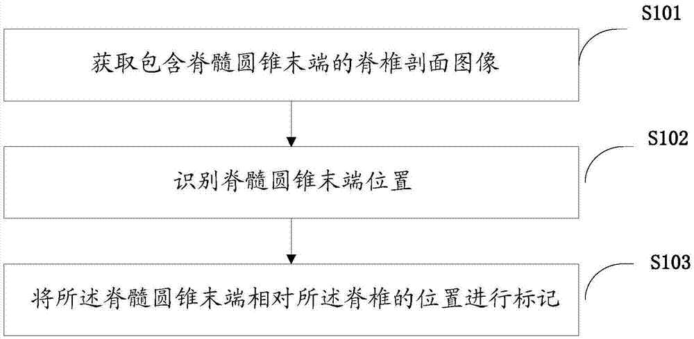
[0034] Such as figure 1 As shown, the present invention provides a kind of target image recognition method, described method comprises the following steps:

[0035] S101. Acquire a cross-sectional image of the spine including the end of the conus medullaris.

[0036] Each vertebra is composed of two vertebral arches and a vertebral body. The spinal cord is located in the character-shaped vertebrae. When the vertebrae are cut open, the low echo dark area inside is the spinal cord. Usually the vertebral body is the best discerned, so the section obtained usually includes at least the vertebral body and the end of the conus medullaris.

[0037] S102, identifying the position of the end of the conus medullaris.

[0038] Since the conus medullaris appears hypoechoic on images, a continuous and narrow hypoechoic region can be obtained after the spine is cut open, so as to further determine the position of the end of the conus medullaris.

[0039] S103. Mark the position of the en...

Embodiment 2

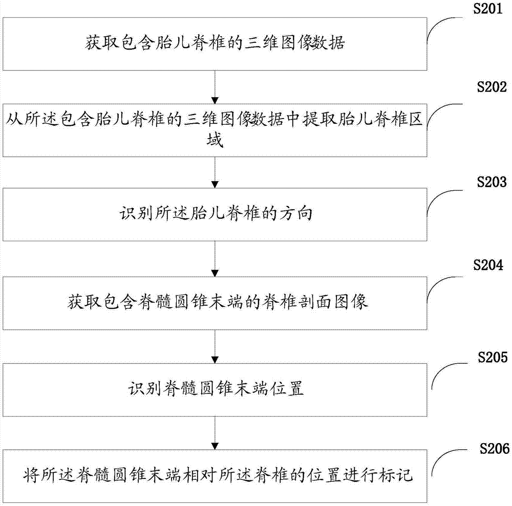
[0103] Such as Figure 5 As shown, according to the above method, the present invention also provides a target image recognition device 300, the device 300 includes: a display unit 301, a storage unit 302, a processor 303; the processor 303 includes: a first image processing A unit 3031, a second image processing unit 3032, and a first control unit 3033;

[0104] The first image processing unit 3031 acquires a spinal section image including the end of the conus medullaris;

[0105] The second image processing unit 3032 identifies the end position of the conus medullaris;

[0106] The first control unit 3033 marks the position of the end of the conus medullaris relative to the spine;

[0107] The display unit 301 is configured to display each ultrasonic image and marker;

[0108] The storage unit 302 is used for storing each ultrasound image and label.

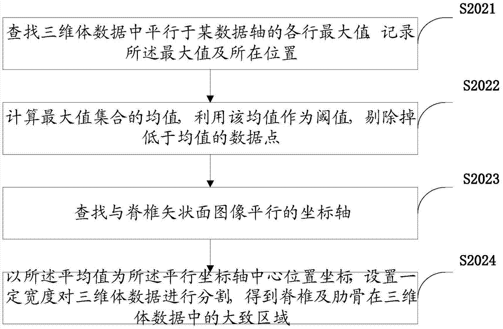
[0109] Such as Image 6 As shown, further, the device 300 further includes: an acquisition unit 3034, a third image proc...

Embodiment 3

[0115] The present invention also provides an ultrasonic imaging device, which includes the device as described in the second embodiment.


PUM


Abstract

The invention provides an automatic target image recognition method. The method comprises the steps of obtaining a spine section image of the tail end of conus medullaris; identifying the position of the tail end of the conus medullaris; marking the position, relative to the spine, of the tail end of the conus medullaris. The invention further provides a corresponding device and ultrasonic equipment thereof. By means of the target image recognition method, the conus medullaris condition of a fetus can be accurately and rapidly diagnosed.

Description

technical field [0001] The invention relates to the technical field of ultrasonic imaging, in particular to a target image recognition method, device and ultrasonic equipment thereof. Background technique [0002] The position of the end of the conus medullaris changes with the growth and development of the fetus: in the first 3 months, the fetal spinal cord is almost as long as the spinal canal, and from the 4th month onwards, the conus medullaris gradually rises relative to the spinal canal. However, if encountering various lesions, the spinal cord can not rise normally, making it lower than the normal level, which may lead to tethered cord. Therefore, prenatal ultrasound positioning of the end of the conus medullaris can provide valuable information for the prenatal diagnosis of fetal spinal region-related diseases, which has important clinical significance. During the examination, the fetus is generally placed in the prone position, and the occipital or parasagittal pla...

Claims


Application Information

Patent Timeline
Patent Type & Authority Applications(China)
IPC IPC(8): A61B8/08G06T7/00
CPCA61B8/085A61B8/0866A61B8/0875A61B8/469A61B8/483A61B8/5207A61B8/5223A61B2576/00G06T7/0012G06T2207/10136G06T2207/30012G06T2207/30044
Inventor 唐艳红董颖艾金钦孙慧李萍
Owner SONOSCAPE MEDICAL CORP
Why Eureka
Try Eureka