
Method and magnetic resonance apparatus for reconstructing an mr image and data carrier

A magnetic resonance equipment and image technology, applied in magnetic resonance measurement, measurement using nuclear magnetic resonance image system, measurement of magnetic variables, etc., can solve problems that affect the quality and effectiveness of MR images

Active Publication Date: 2016-03-30
SIEMENS HEALTHCARE GMBH
View PDF5 Cites 6 Cited by
  • Summary
  • Abstract
  • Description
  • Claims
  • Application Information

AI Technical Summary

Problems solved by technology

However, it is at high field strengths that, for example, chemical shifts between fat and water cause artifacts (so-called CSD artifacts (“Chemical Shift Displacement”, chemical shifts)), which negatively affect the quality of the created MR images. Image Quality and Availability

Method used


Image

  • Method and magnetic resonance apparatus for reconstructing an mr image and data carrier
  • Method and magnetic resonance apparatus for reconstructing an mr image and data carrier
  • Method and magnetic resonance apparatus for reconstructing an mr image and data carrier

Examples


Embodiment Construction

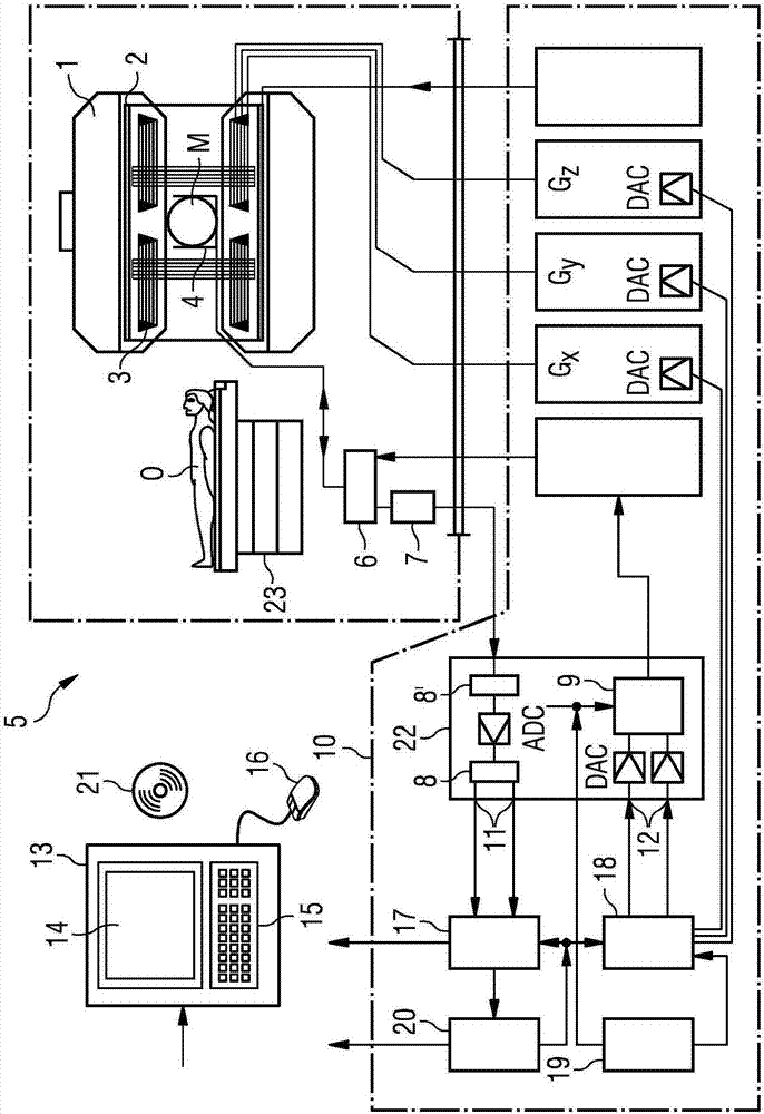
[0049] figure 1 A magnetic resonance device 5 (magnetic resonance imaging or nuclear magnetic resonance imaging device ( )) schematic diagram. Here, the main field magnet 1 generates a temporally constant magnetic field for polarizing or aligning an object O, for example a core rotation in the examination region of a part to be examined of a human body lying flat on a table 23 which is continuously pushed into the magnetic resonance apparatus 5. The high homogeneity of the main magnetic field necessary for nuclear magnetic resonance measurements is defined in a typically spherical measurement volume M in which the part of the human body to be examined is preferably measured. In order to support the homogeneity requirements and in particular to eliminate time-invariant influences, so-called shim laminations consisting of ferromagnetic elements are installed at suitable points. Temporal variations are eliminated by shim coils 2 .

[0050] A cylindrical gradient field system...


PUM


Abstract

In a method and magnetic resonance apparatus (5) for reconstructing an MR image (43) of a volume segment within an examination object (O), MR data within the volume segment are acquired according to a separation method by MR data for a k-space line being acquired respectively in steps while a readout gradient is activated in the same readout direction. An image (41) of a first substance and an image (42) of a second substance are thereby reconstructed. A shift length (V), by which the image of the first substance and the image of the second substance are displaced relative to one another due to the chemical shift in the readout direction is determined. The image of the first substance and / or the image of the second substance is shifted as a function of the shift length, to compensate for the relative displacement between the images of the first and second substances in the readout direction due to the chemical shift. An MR image is generated by combining the images of the first and second substances. A data carrier is also provided.

Description

technical field [0001] The invention relates to a method and a magnetic resonance system as well as a data carrier for reconstructing MR images in which chemical shifts are taken into account. Background technique [0002] For example, in orthopedic imaging, MR images are required for diagnosis, in which no fat suppression was used during the setup. Furthermore, high resolution and high signal-to-noise ratios are sought in orthopedic imaging, which can only be achieved with high field strengths (eg three Tesla or higher). However, it is at high field strengths that, for example, chemical shifts between fat and water cause artifacts (so-called CSD artifacts (“Chemical Shift Displacement”, chemical shifts)), which negatively affect the quality of the created MR images. Image quality and availability. Contents of the invention [0003] It is therefore the object of the present invention to create an MR image in which a plurality of materials (for example fat and water) can ...

Claims


Application Information

Patent Timeline
Patent Type & Authority Applications(China)
IPC IPC(8): G01R33/56A61B5/055G01R33/485
CPCG01R33/56527G01R33/4818G01R33/4828
Inventor 多米尼克·保罗本杰明·施米特
Owner SIEMENS HEALTHCARE GMBH
Learn More