Method and system for inhibiting bone shadows in digital X ray chest radiograph

An image center and bone technology, applied in the field of image processing, can solve the problems of increased radiation dose received by patients, achieve the effect of enhancing visibility and solving the overlap of anatomical structure images

Active Publication Date: 2016-04-06
深圳市深图医学影像设备有限公司
View PDF5 Cites 5 Cited by
  • Summary
  • Abstract
  • Description
  • Claims
  • Application Information

AI Technical Summary

Problems solved by technology

[0004] The technical problem to be solved by the present invention is to provide a method and system for suppressing bone shadows in digitized chest X-ray images, aiming to solve the problem that shadow suppression of chest X-ray images can only be realized under the dual-energy subtraction technology in the prior art. Problems causing increased radiation doses to patients

Method used


Image

  • Method and system for inhibiting bone shadows in digital X ray chest radiograph
  • Method and system for inhibiting bone shadows in digital X ray chest radiograph
  • Method and system for inhibiting bone shadows in digital X ray chest radiograph

Examples


Embodiment Construction

[0072] In order to make the object, technical solution and advantages of the present invention clearer, the present invention will be further described in detail below in conjunction with the accompanying drawings and embodiments. It should be understood that the specific embodiments described here are only used to explain the present invention, not to limit the present invention.

[0073] The invention provides a method and system for suppressing bone images in digitized X-ray images without X-ray dual-energy subtraction photography. Specifically, the present invention suppresses images of ribs and clavicles in a single digitized chest X-ray taken by common X-ray DR or CR equipment.

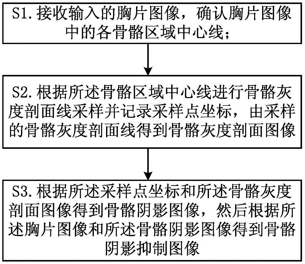
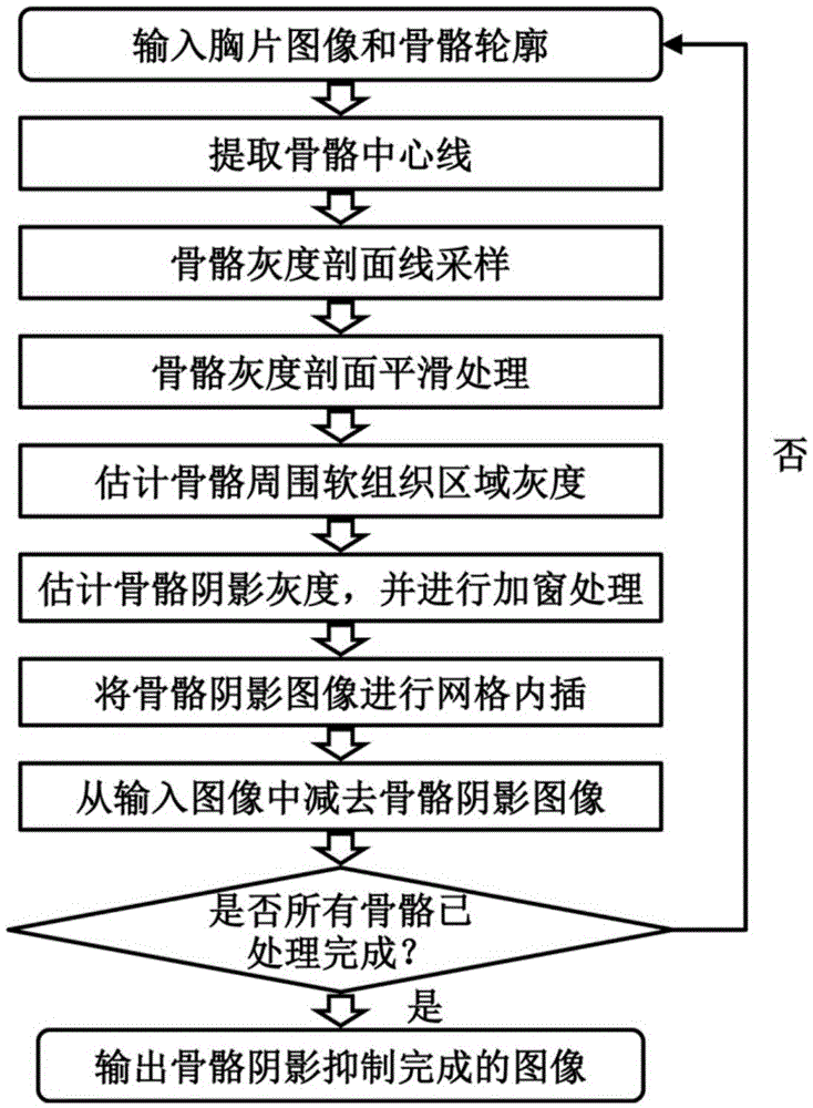
[0074] Therefore, if figure 1 As shown, a method for suppressing bone shadows in a digitized X-ray chest image provided by Embodiment 1 of the present invention, the steps include:

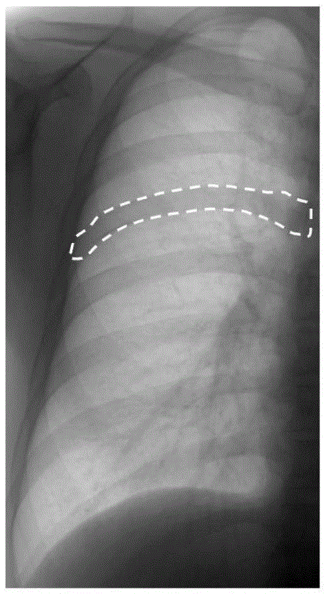
[0075] S1, receiving the input chest radiograph image, and confirming the centerline of each bone region in the...


PUM


Abstract

The invention is suitable for the field of image processing and provides a method for inhibiting bone shadows in a digital X ray chest radiograph. The method comprises the steps of: A, receiving an input chest radiograph, and determining a central line of each bone area in the chest radiograph; B, according to the central lines of the bone areas, carrying out bone gray section line sampling and recording coordinates of sampling points, and obtaining a bone gray section image by the sampled bone gray section lines; and C, according to the coordinates of sampling points and the bone gray section image, obtaining a bone shadow image, and then according to the chest radiograph and the bone shadow image, obtaining bone shadow inhibited image. According to the invention, an X ray dual energy subtraction device is not needed, and modeling learning using a dual energy subtraction image is not needed, the single general ray chest radiograph is processed, and bone shadow inhibition is realized for the chest radiograph; and furthermore, a smooth and curved surface fitting technology is utilized to estimate the bone shadow gray, and detailed structures in the input X ray chest radiograph can be effectively maintained.

Description

technical field [0001] The invention belongs to the field of image processing, in particular to a method and system for suppressing bone shadows in digital X-ray chest images. Background technique [0002] Chest X-ray plain film (abbreviated as chest film) is the basic imaging method for detecting pulmonary nodules and diagnosing lung diseases. However, the anatomical structures overlap each other in conventional chest radiographs, and some small nodules and lesions in the lungs may be missed due to the occlusion of the ribs and clavicles. Detection of nodules obscured by ribs and clavicles is also difficult for some computer-aided diagnosis software on chest radiographs. Suppressing bone images in chest X-rays can solve the problem of image overlap of anatomical structures, enhance the visibility of X-ray chest X-rays, and facilitate doctors to perform image interpretation and diagnosis and computer-aided diagnosis software for processing. [0003] Dual Energy Subtraction...

Claims


Application Information

Patent Timeline
Patent Type & Authority Applications(China)
IPC IPC(8): G06T5/00
CPCG06T5/002G06T2207/10116G06T2207/20182G06T2207/30061G06T2207/30064
Inventor 饶玉明阳维王益民
Owner 深圳市深图医学影像设备有限公司
Why Eureka
Try Eureka